

Matrix metalloproteinases, new insights into the understanding of neurodegenerative disorders
- PMID: 24116286
- PMCID: PMC3792209
- DOI: 10.4062/biomolther.2012.20.2.133
Matrix metalloproteinases, new insights into the understanding of neurodegenerative disorders
Abstract
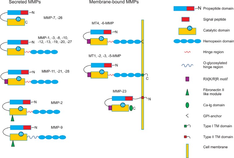
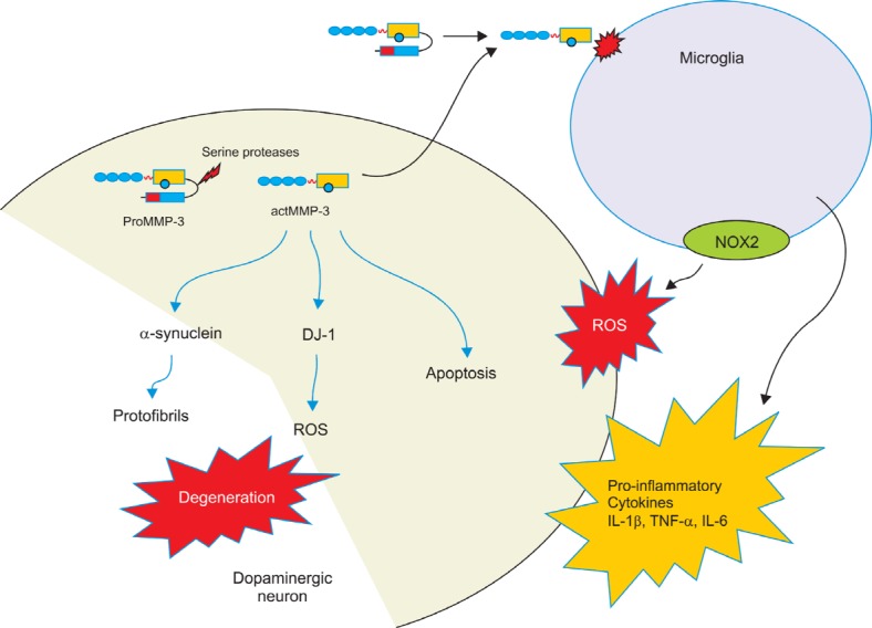
Matrix metalloproteinases (MMPs) are a subfamily of zinc-dependent proteases that are responsible for degradation and remodeling of extracellular matrix proteins. The activity of MMPs is tightly regulated at several levels including cleavage of prodomain, allosteric activation, compartmentalization and complex formation with tissue inhibitor of metalloproteinases (TIMPs). In the central nervous system (CNS), MMPs play a wide variety of roles ranging from brain development, synaptic plasticity and repair after injury to the pathogenesis of various brain disorders. Following general discussion on the domain structure and the regulation of activity of MMPs, we emphasize their implication in various brain disorder conditions such as Alzheimer's disease, multiple sclerosis, ischemia/reperfusion and Parkinson's disease. We further highlight accumulating evidence that MMPs might be the culprit in Parkinson's disease (PD). Among them, MMP-3 appears to be involved in a range of pathogenesis processes in PD including neuroinflammation, apoptosis and degradation of α-synuclein and DJ-1. MMP inhibitors could represent potential novel therapeutic strategies for treatments of neurodegenerative diseases.
Keywords: MMP-3; Matrix metalloproteinases; Microglia; Neurodegenerative disorders; Parkinson’s disease.
Figures
Similar articles
-
Multifaceted Role of Matrix Metalloproteinases in Neurodegenerative Diseases: Pathophysiological and Therapeutic Perspectives.Int J Mol Sci. 2021 Jan 30;22(3):1413. doi: 10.3390/ijms22031413. Int J Mol Sci. 2021. PMID: 33573368 Free PMC article. Review.
-
Implication of matrix metalloproteinases in regulating neuronal disorder.Mol Biol Rep. 2015 Jan;42(1):1-11. doi: 10.1007/s11033-014-3752-y. Mol Biol Rep. 2015. PMID: 25374425 Review.
-
Matrix metalloproteinases and their multiple roles in neurodegenerative diseases.Lancet Neurol. 2009 Feb;8(2):205-16. doi: 10.1016/S1474-4422(09)70016-X. Lancet Neurol. 2009. PMID: 19161911 Review.
-
Multifaceted role of matrix metalloproteinases (MMPs).Front Mol Biosci. 2015 May 13;2:19. doi: 10.3389/fmolb.2015.00019. eCollection 2015. Front Mol Biosci. 2015. PMID: 25988186 Free PMC article. Review.
-
[Matrix metalloproteinases and their inhibitors, modulators of neuro-immune interactions and of pathophysiological processes in the nervous system].J Soc Biol. 2003;197(2):133-44. J Soc Biol. 2003. PMID: 12910628 Review. French.
Cited by
-
A new triazine bearing a pyrazolone group capable of copper, nickel, and zinc chelation.RSC Adv. 2018;8(6):3024-3035. doi: 10.1039/c7ra09459k. Epub 2018 Jan 16. RSC Adv. 2018. PMID: 29503730 Free PMC article.
-
Human Papillomavirus Modulates Matrix Metalloproteinases During Carcinogenesis: Clinical Significance and Role of Viral Oncoproteins.In Vivo. 2022 Nov-Dec;36(6):2531-2541. doi: 10.21873/invivo.12990. Epub 2022 Oct 29. In Vivo. 2022. PMID: 36309355 Free PMC article. Review.
-
An Iatrogenic Model of Brain Small-Vessel Disease: Post-Radiation Encephalopathy.Int J Mol Sci. 2020 Sep 5;21(18):6506. doi: 10.3390/ijms21186506. Int J Mol Sci. 2020. PMID: 32899565 Free PMC article. Review.
-
Sijunzi decoction may decrease apoptosis via stabilization of the extracellular matrix following cerebral ischaemia-reperfusion in rats.Exp Ther Med. 2019 Oct;18(4):2805-2812. doi: 10.3892/etm.2019.7878. Epub 2019 Aug 13. Exp Ther Med. 2019. PMID: 31572528 Free PMC article.
-
Microglial interactions with the neurovascular system in physiology and pathology.Dev Neurobiol. 2018 Jun;78(6):604-617. doi: 10.1002/dneu.22576. Epub 2018 Feb 1. Dev Neurobiol. 2018. PMID: 29318762 Free PMC article. Review.
References
-
- Agrawal S. Agrawal P. Durbeej M. van Rooijen N. Ivars F. Opdenakker G. Sorokin L. M. Dystroglycan is selectively cleaved at the parenchymal basement membrane at sites of leukocyte extravasation in experimental autoimmune encephalomyelitis. J. Exp. Med. (2006);203:1007–1019. doi: 10.1084/jem.20051342. - DOI - PMC - PubMed
-
- Asahi M. Asahi K. Jung J. C. del Zoppo G. J. Fini M. E. Lo E. H. Role for matrix metalloproteinase 9 after focal cerebral ischemia: effects of gene knockout and enzyme inhibition with BB-94. J. Cereb. Blood Flow Metab. (2000);20:1681–1689. - PubMed
Publication types
LinkOut - more resources
Full Text Sources
Other Literature Sources
Miscellaneous
