

Cerebral vasculitis in adults: what are the steps in order to establish the diagnosis? Red flags and pitfalls
- PMID: 24117125
- PMCID: PMC3927902
- DOI: 10.1111/cei.12221
Cerebral vasculitis in adults: what are the steps in order to establish the diagnosis? Red flags and pitfalls
Abstract
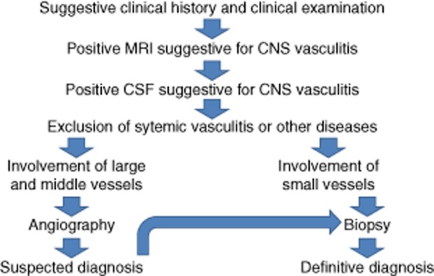
Cerebral vasculitis is a rare cause of juvenile stroke. It may occur as primary angiitis of the central nervous system (PACNS) or as CNS manifestation in the setting of systemic vasculitis. Clinical hints for vasculitis are headache, stroke, seizures, encephalopathy and signs of a systemic inflammatory disorder. Diagnostic work-up includes anamnesis, whole body examination, laboratory and cerebral spinal fluid (CSF) studies, magnetic resonance imaging (MRI), angiography and brain biopsy. Due to the rarity of the disease, exclusion of more frequent differential diagnoses is a key element of diagnostic work -up. This review summarizes the steps that lead to the diagnosis of cerebral vasculitis and describes the red flags and pitfalls. Despite considering the dilemma of angiography-negative vasculitis and false-negative brain biopsy in some cases, it is important to protect patients from 'blind' immunosuppressive therapy in unrecognized non-inflammatory differential diagnosis.
Keywords: encephalitis; granulomatosis with polyangiitis; stroke; vasculitis.
© 2013 British Society for Immunology.
Figures
References
-
- Berlit P. Primary angiitis of the CNS – an enigma that needs world-wide efforts to be solved. Eur J Neurol. 2009;16:10–11. - PubMed
-
- Neel A, Auffray-Calvier E, Guillon B, et al. Challenging the diagnosis of primary angiitis of the central nervous system: a single-center retrospective study. J Rheumatol. 2012;39:1026–1034. - PubMed
-
- Lopez JI, Holdridge A, Chalela J. Headache and vasculitis. Curr Pain Headache Rep. 2013;17:320. - PubMed
-
- Kramer M, Berlit P. [Reversible cerebral vasoconstriction syndrome vs cerebral vasculitis? On the importance and difficulty of differentiating] Nervenarzt. 2011;82:500–505. - PubMed
-
- Singhal AB, Hajj-Ali RA, Topcuoglu MA, et al. Reversible cerebral vasoconstriction syndromes: analysis of 139 cases. Arch Neurol. 2011;68:1005–1012. - PubMed
Publication types
MeSH terms
LinkOut - more resources
Full Text Sources
Other Literature Sources
