. 2013 Oct 23;135(42):15726-9.
doi: 10.1021/ja408760w. Epub 2013 Oct 11.

DNA protection by the bacterial ferritin Dps via DNA charge transport

Affiliations

DNA protection by the bacterial ferritin Dps via DNA charge transport

Anna R Arnold et al. J Am Chem Soc. .

Abstract

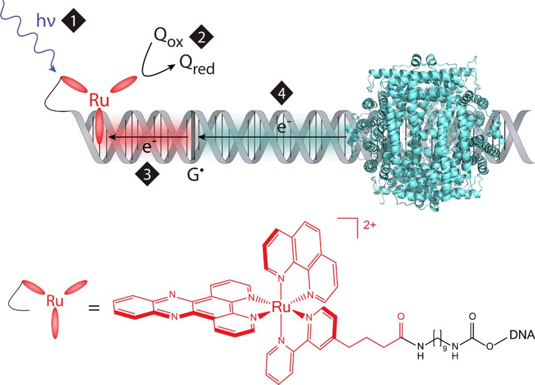
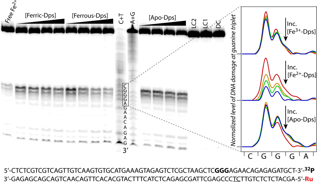
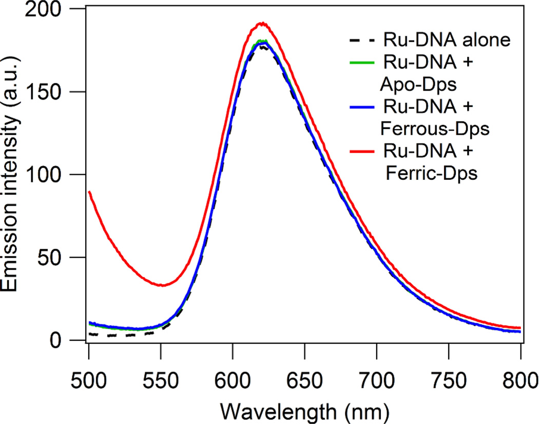
Dps proteins, bacterial mini-ferritins that protect DNA from oxidative stress, are implicated in the survival and virulence of pathogenic bacteria. Here we examine the mechanism of E. coli Dps protection of DNA, specifically whether this DNA-binding protein can utilize DNA charge transport through the base pair π-stack to protect the genome from a distance. An intercalating ruthenium photooxidant was employed to generate DNA damage localized to guanine repeats, the sites of lowest potential in DNA. We find that Dps loaded with ferrous iron, in contrast to Apo-Dps and ferric iron-loaded Dps, significantly attenuates the yield of oxidative DNA damage. These data demonstrate that ferrous iron-loaded Dps is selectively oxidized to fill guanine radical holes, thereby restoring the integrity of the DNA. Luminescence studies indicate no direct interaction between the ruthenium photooxidant and Dps, supporting the DNA-mediated oxidation of ferrous iron-loaded Dps. Thus DNA charge transport may be a mechanism by which Dps efficiently protects the genome of pathogenic bacteria from a distance.


Figures

Figure 1
Figure 1
Schematic depicting DNA-mediated oxidation of Dps to fill the guanine radical hole generated by flash-quench chemistry. Upper: First, visible light excites an intercalated ruthenium(II) photooxidant (1), which is then oxidatively quenched (2) to a highly oxidizing Ru(III) species by a diffusing quencher (Q). This Ru(III) species is competent to abstract an electron from DNA (3); the hole equilibrates along the π-stack and localizes to the 5’-G of guanine repeats, the most easily oxidized base (G•). DNA CT from Dps to the guanine radical (4) could be a long distance protection mechanism. Note that the location and precise geometry for DNA binding of Dps are unknown. E. coli Dps PDB: 1dps. Lower: Structure of [Ru(phen)(dppz)(bpy’)]2+ covalently tethered to DNA via diaminononane linkage.
Figure 2
Figure 2
Representative gel comparing ability of Apo-Dps, ferrous-iron loaded Dps, and ferric-iron loaded Dps to protect DNA from damage created via flash-quench chemistry. Left: Autoradiogram of gel showing effect of increasing Dps with varying iron loading. Conditions: 6 µM statistically 3’-32P-labeled 70 mer Ru-DNA, 600 µM [Co(NH3)5Cl]2+, Dps concentrations from 0 to 6 µM, buffer: 50 mM Tris, pH 7.0, 150 mM NaCl. A+G and C+T are Maxam-Gilbert sequencing lanes. Controls: Dark control (DC) contains all components (DNA, photooxidant, and quencher) but is not irradiated; Light control 1 (LC1) is irradiated but lacks quencher; LC2 is irradiated, lacking quencher, but contains 6 µM Dps loaded with ferric iron; Free Fe2+ is irradiated, containing all components but protein, with the addition of free ferrous iron at a concentration equivalent to that in ferrous iron-loaded Dps. The number of Fe/Dps in ferrous iron-loaded Dps was 10.8 ± 0.1. Right: Lane profiles comparing normalized level of DNA damage at the guanine triplet upon titration of ferric iron-loaded Dps (upper), ferrous iron-loaded Dps (middle), or Apo-Dps (lower) from 0 µM protein (red), 2 µM protein (orange), 4 µM protein (green), to 6 µM protein (blue). Lower: DNA sequence, with guanine triplet shown bolded, and the location of a nick in the DNA backbone underlined. For higher synthetic yield, the DNA was made in two pieces (55-mer and Ru-15-mer); breaks in the sugar-phosphate backbone do not affect long-range CT.
Figure 3
Figure 3
Ru-DNA luminescence in absence and presence of Dps. Anaerobically prepared samples containing 8 µM duplexed 70-mer DNA with covalently tethered [Ru(phen)(dppz)(bpy’)]2+ alone or with 8 µM Apo-Dps, ferrous-iron loaded Dps (11.8 ± 0.2 Fe/Dps dodecamer), or ferric-iron loaded Dps in 50 mM Tris, pH 7.0, 150 mM NaCl. Excitation wavelength: 440 nm. Slight precipitation occurs for the sample containing Ru-DNA and ferric iron-loaded Dps, resulting in a raised baseline.

References

    1. Zeth K. Biochem. J. 2012;445:297–311. - PubMed
    1. Ceci P, Cellai S, Falvo E, Rivetti C, Rossi GL, Chiancone E. Nucleic Acids Res. 2004;32:5935–5944. - PMC - PubMed
    1. Sund CJ, Rocha ER, Tzinabos AO, Wells WG, Gee JM, Reott MA, O’Rourke DP, Smith CJ. Mol. Microbiol. 2008;67:129–142. - PubMed
    1. Li X, Pal U, Ramamoorthi N, Liu X, Desrosiers DC, Eggers CH, Anderson JF, Radolf JD, Fikrig E. Mol. Microbiol. 2007;63:694–710. - PubMed
    1. (a) Helicobacter pylori (neutrophil-activating protein, HP-NAP) D’Elios MM, Amedei A, Cappon A, Del Prete G, de Bernard M. FEMS Immunol. Med. Mic. 2007;50:157–164. (b) Salmonella enterica serovar Typhimurium Halsey TA, Vazquez-Torres A, Gravdahl DJ, Fang FC, Libby SJ. Infect. Immun. 2004;72:1155–1158. and (c) Porphyromonas gingivalis Ueshima J, Shoji M, Ratnayake DB, Abe K, Yoshida S, Yamamoto K, Nakayama K. Infect. Immun. 2003;71:1170–1178.

Publication types

MeSH terms

LinkOut - more resources