Review
. 2013 Nov;256(1):148-59.
doi: 10.1111/imr.12120.

Regulation of T-cell receptor signaling by the actin cytoskeleton and poroelastic cytoplasm

Affiliations
Review

Regulation of T-cell receptor signaling by the actin cytoskeleton and poroelastic cytoplasm

Peter Beemiller et al. Immunol Rev. 2013 Nov.

Abstract

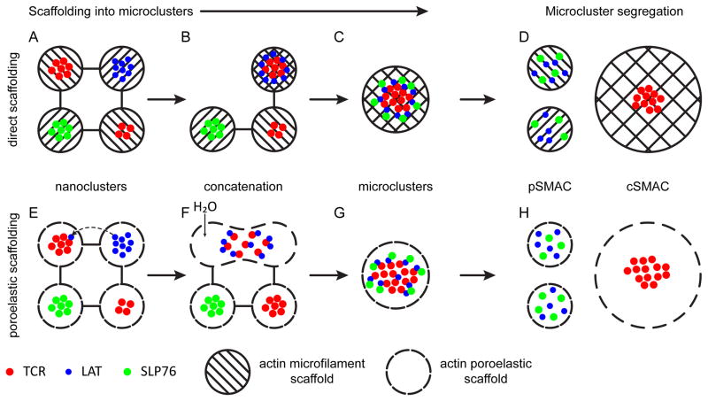
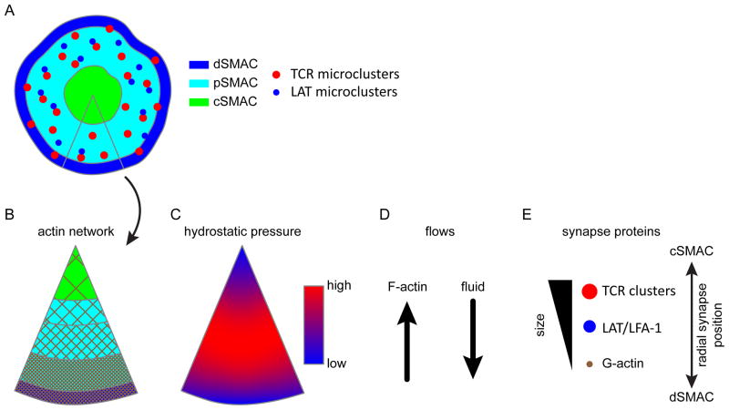
The actin cytoskeleton plays essential roles in modulating T-cell activation. Most models of T-cell receptor (TCR) triggering signalosome assembly and immune synapse formation invoke actin-dependent mechanisms. As T cells are constitutively motile cells, TCR triggering and signaling occur against a cytoskeletal backdrop that is constantly remodeling. While the interplay between actin dynamics and TCR signaling have been the focus of research for many years, much of the work in T cells has considered actin largely for its 'scaffolding' function. We examine the roles of the actin cytoskeleton in TCR signaling and immune synapse formation with an emphasis on how poroelasticity, an ensemble feature of actin dynamics with the cytosol, relates to how T cells respond to stimulation.

Keywords: T cells; T-cell receptors; signal transduction.


Conflict of interest statement

The authors declare that they have no financial conflict of interest.

Figures

Fig. 1
Fig. 1. Models of T-cell receptor scaffolding
Two models of signaling microcluster scaffolding. (A–D) A direct, microfilament interaction-based model of signalosome scaffolding. (E–H) A poroelastic model of signalosome scaffolding. (A) TCRs and signaling effectors like LAT interact with the actin cytoskeleton, organizing into nanoclusters. (B) Upon triggering, actin cytoskeletal rearrangements allow protein islands of TCRs and LAT to concatenate. (C) Further concatenation and cytoskeletal remodeling leads to the aggregation of microclusters and the recruitment of other signaling effectors, such as SLP-76. (D) Association with signaling effectors is dependent on the actin cytoskeleton for scaffolding. As a result, as microclusters move into the cSMAC, signaling effectors are lost from TCR microclusters. In the pSMAC, the effectors can be rescaffolded with F-actin. (E) The actin cytoskeleton corrals TCRs into cytosolic pores. Confinement to a pore is not absolute, and diffusion into nearby pores can occur (dotted line). The rate at which molecules move into neighboring pores would be inversely proportional to the strength of the homotypic interactions holding molecules in nanoclusters and the hydrodynamic radius of the molecule. (F) As signaling is initiated, water influx, possibly induced by Na-H antiporter activity, changes the local hydrostatic pressure, causing pore deformation and swelling. This increases the mean pore size, allowing nanoclusters of TCRs and LAT to merge in growing pores. (G) Continued hydrostatic pressure facilitates the merging of TCR nanoclusters into microclusters and the incorporation of more signaling effector molecules, such as SLP-76 to sustain signaling. (H) In the low F-actin density interior of the synapse, effectors that require actin scaffolding to remain associated with TCRs release and are separated from TCRs by anterograde fluid flow. The molecules are free to diffuse back into smaller peripheral pores, terminating TCR signaling. Meanwhile, TCRs, which have become independent of the actin cytoskeleton for association in microclusters, remain trapped in the cSMAC. The TCRs cannot diffuse into the periphery of the synapse due to the fine pore size of the pSMAC.
Fig. 2
Fig. 2. Models of cellular transport
(A) The organization of the immune synapse macromolecular domains. Microclusters are generated in the periphery in the pSMAC and dSMAC, and then flow into the cSMAC. (B) A slice through the immune synapse domains showing the branched actin network. F-actin density is highest in the peripheral SMAC zones. As a result, the cytosolic pore size is inversely proportional to the distance from the center of the synapse. Due to the small pore size in the pSMAC, signaling complexes are efficiently coupled to retrograde actin flow. (C) A proposed, simplified map of hydrostatic pressure across the synapse. (D) As signaling complexes centralize, they are simultaneously subjected to retrograde actin flow toward the cSMAC and anterograde fluid flow through the poroelastic media toward the periphery of the cell. (E) The two forces counteract each other, modulating the flow of solid material through the cell in a size-dependent manner. The balance of these two forces regulates the speed and direction of protein complex movement. Large complexes, such as TCRs microclusters, segregate into the interior region with a larger cytosolic pore size. Intermediate size complexes, such as LAT and integrin microclusters, are coupled to actin retrograde flow in the pSMAC but are unstable in the cSMAC. Very small particles, such as actin monomers, are driven by hydrostatic pressure to the edge of the synapse as in migrating cells, allowing continuous actin treadmilling.

References

    1. Valitutti S, Dessing M, Aktories K, Gallati H, Lanzavecchia A. Sustained signaling leading to T cell activation results from prolonged T cell receptor occupancy. Role of T cell actin cytoskeleton. J Exp Med. 1995;181:577–584. - PMC - PubMed
    1. Campi G, Varma R, Dustin ML. Actin and agonist MHC-peptide complex-dependent T cell receptor microclusters as scaffolds for signaling. J Exp Med. 2005;202:1031–1036. - PMC - PubMed
    1. Huppa JB, Axmann M, Mörtelmaier MA, Lillemeier BF, Newell EW, Brameshuber M, Klein LO, et al. TCR-peptide-MHC interactions in situ show accelerated kinetics and increased affinity. Nature. 2010;463:963–967. - PMC - PubMed
    1. Huang J, Zarnitsyna VI, Liu B, Edwards LJ, Jiang N, Evavold BD, Zhu C. The kinetics of two-dimensional TCR and pMHC interactions determine T-cell responsiveness. Nature. 2010;464:932–936. - PMC - PubMed
    1. Valitutti S, Muller S, Cella M, Padovan E, Lanzavecchia A. Serial triggering of many T-cell receptors by a few peptide–MHC complexes. Nature. 1995;375:148–151. - PubMed

Publication types

Substances