. 2014 Feb;16(2):247-68.
doi: 10.1111/cmi.12219. Epub 2013 Nov 3.

Yersinia pseudotuberculosis uses Ail and YadA to circumvent neutrophils by directing Yop translocation during lung infection

Affiliations

Yersinia pseudotuberculosis uses Ail and YadA to circumvent neutrophils by directing Yop translocation during lung infection

Michelle K Paczosa et al. Cell Microbiol. 2014 Feb.

Abstract

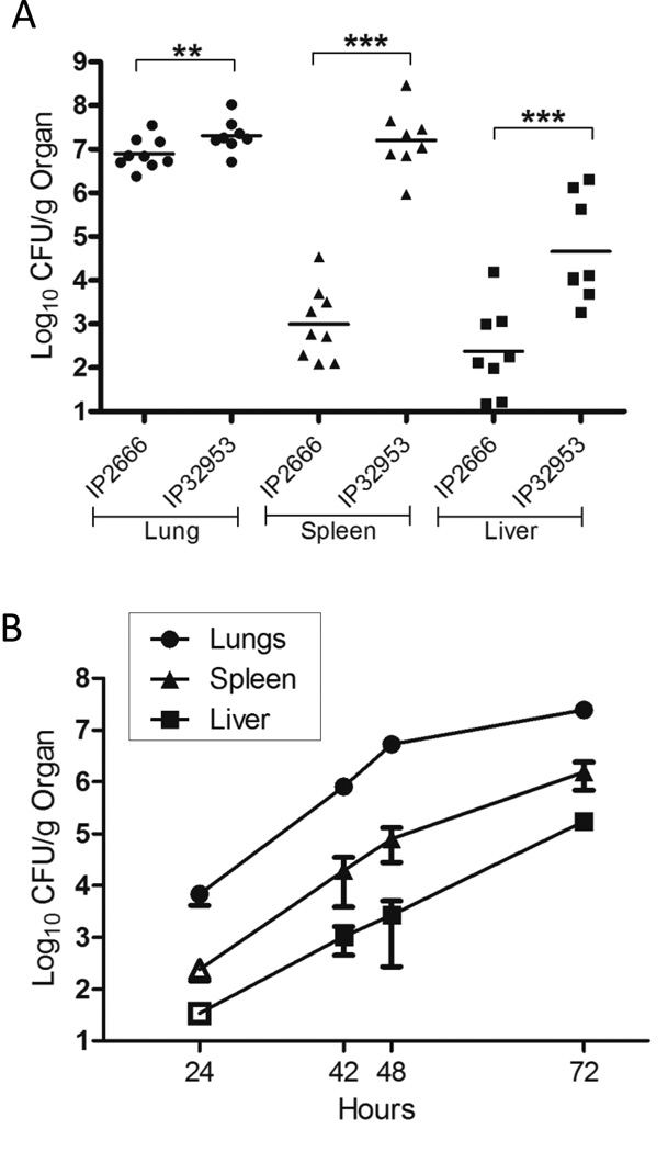
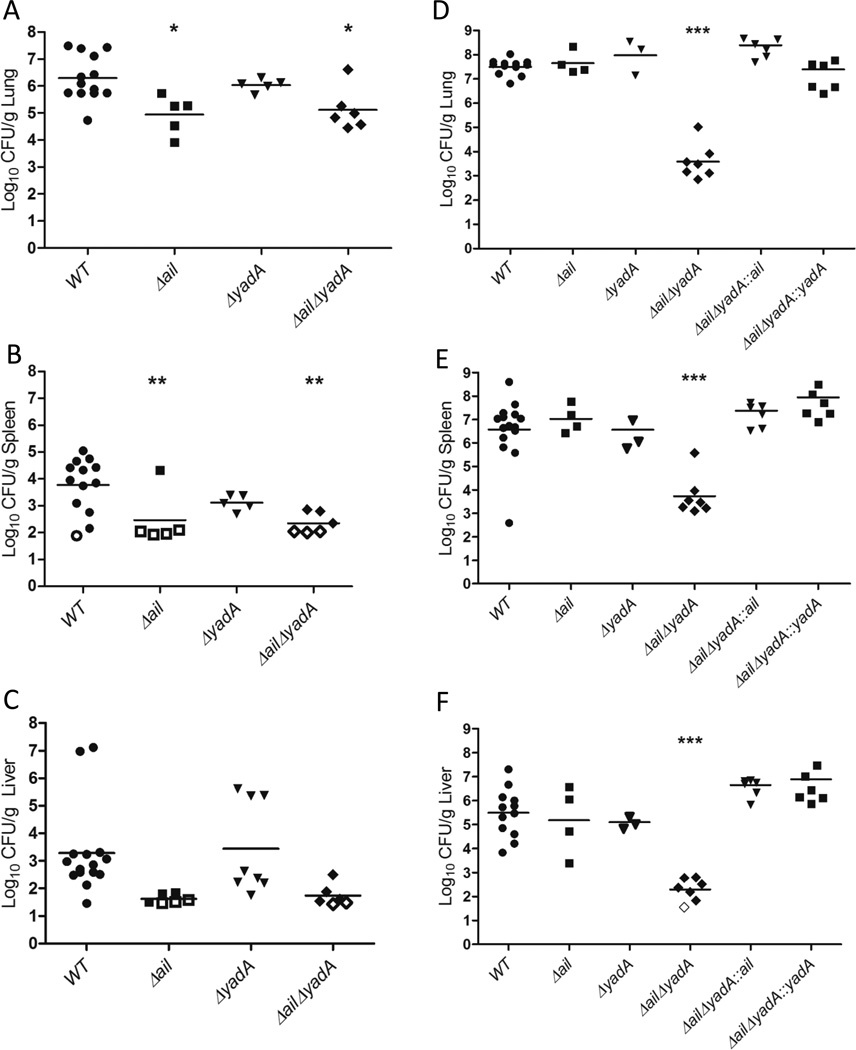
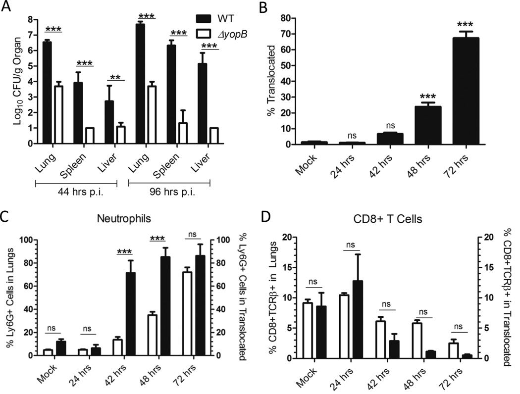
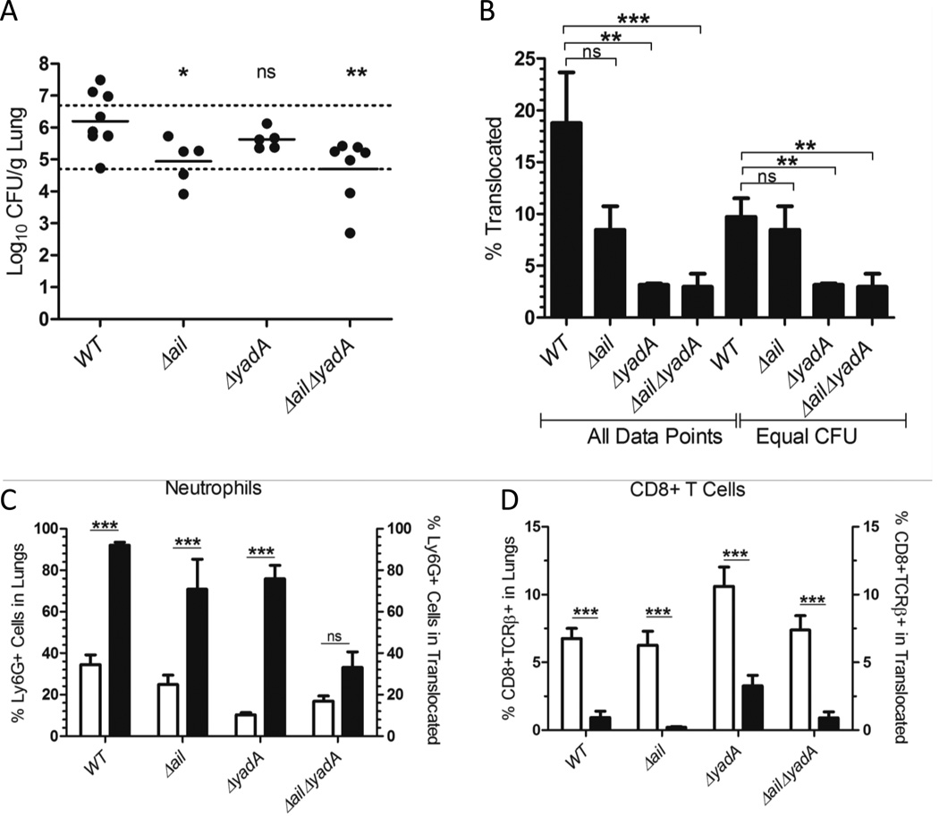
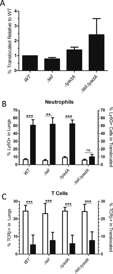
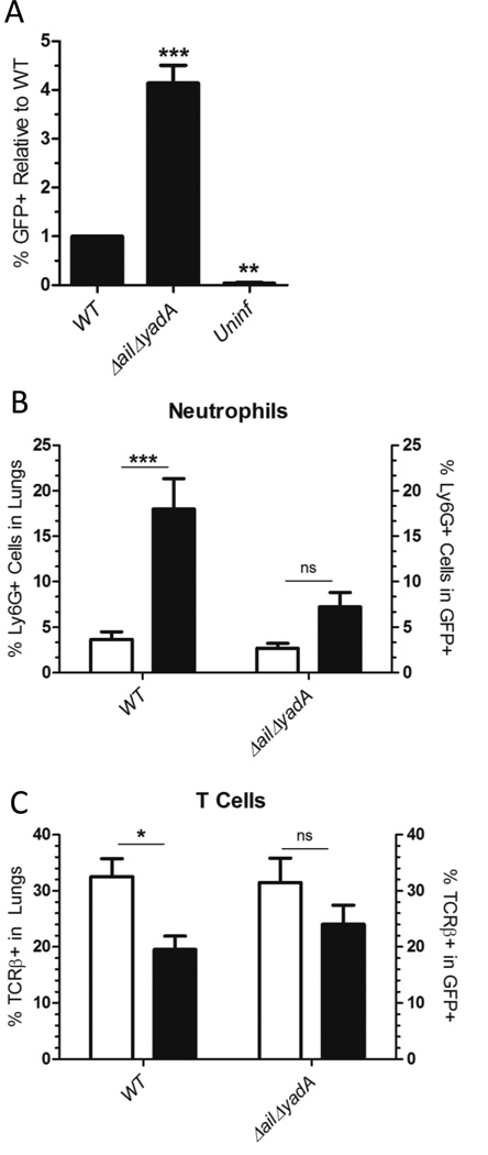
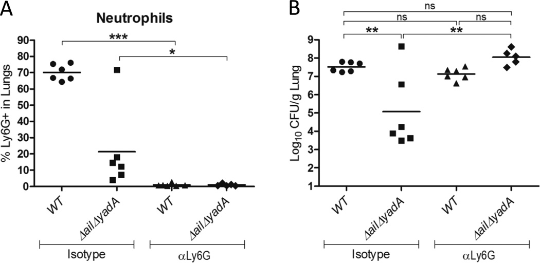
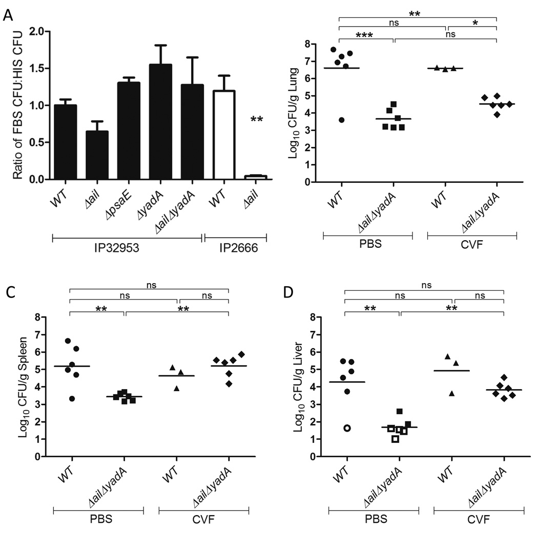
A Yersinia pseudotuberculosis (Yptb) murine model of lung infection was previously developed using the serotype III IP2666NdeI strain, which robustly colonized lungs but only sporadically disseminated to the spleen and liver. We demonstrate here that a serotype Ib Yptb strain, IP32953, colonizes the lungs at higher levels and disseminates more efficiently to the spleen and liver compared with IP2666NdeI . The role of adhesins was investigated during IP32953 lung infection by constructing isogenic Δail, Δinv, ΔpsaE and ΔyadA mutants. An IP32953ΔailΔyadA mutant initially colonized but failed to persist in the lungs and disseminate to the spleen and liver. Yptb expressing these adhesins selectively bound to and targeted neutrophils for translocation of Yops. This selective targeting was critical for virulence because persistence of the ΔailΔyadA mutant was restored following intranasal infection of neutropenic mice. Furthermore, Ail and YadA prevented killing by complement-mediated mechanisms during dissemination to and/or growth in the spleen and liver, but not in the lungs. Combined, these results demonstratethat Ail and YadA are critical, redundant virulence factors during lung infection, because they thwart neutrophils by directing Yop-translocation specifically into these cells.


Figures

Figure 1
Figure 1. IP32953 disseminates more rapidly than IP2666 following intranasal infection
(A) BALB/c mice were intranasally infected with (A) 500 CFU of the WT IP2666NdeI or WT IP32953 strains, or (B) 250 CFU of WT IP32953. (A) 96 h.p.i. lungs (circles), spleens (triangles), and livers (squares) were harvested and plated for CFU. Data is representative of at least two independent experiments with 2–4 mice per experiment, Each dot represents an individual mouse. The bars represent the geometric mean and the asterisks represent significance as analyzed by Student’s T-test (**p<0.01, ***p<0.001). (B) At time points indicated, lungs (circles), spleens (triangles) and livers (squares) were harvested and plated for CFU. Open symbol indicates the limit of detection and that no bacteria were recovered at that time-point in any mice. The error bars are the standard error of the mean (SEM). Each point is the mean from 3 independent experiments consisting of 3 mice per time point per experiment.
Figure 2
Figure 2. Ail is critical for early lung colonization and dissemination to the spleen and liver, while Ail and YadA are redundant and critical for IP32953 persistence in the lungs
BALB/c mice were infected intranasally with 250 CFU of the indicated IP32953 strain. At (A–C) 44 h.p.i. or (D–F) 96 h.p.i., the (A, D) lungs, (B, E) spleens, and (C, F) livers were harvested and plated for CFU. Each dot represents the CFU recovered from an individual mouse, and open symbols indicate that no bacteria were recovered at that limit of detection. The bars represent the geometric mean. (A-C) 2–4 independent experiments of 2 or 3 mice per strain were performed. (D–F) 1–4 independent experiments/strain of 2–3 mice per experiment were performed. Significance was determined by One-Way ANOVA with Tukey’s post-test. Asterisks indicate significance of mutants as compared to WT values (*p<0.05, **p<0.01, ***p<0.001).
Figure 3
Figure 3. Yop translocation by IP32953 is critical for lung colonization and dissemination to the spleen and liver, and is targeted preferentially into neutrophils during lung infection
(A) BALB/c mice were infected intranasally with 250 CFU of IP32953 WT (black) or ΔyopB (grey). At 44 and 96 h.p.i., the lungs, spleens and livers were harvested and plated for CFU. Data is from 2 independent experiments with 3 mice per strain per experiment. The log10 mean of the CFU and SEM are shown. Statistical significance was determined by One-Way ANOVA with Tukey’s post-test. (B–D) BALB/c mice were infected intranasally with 250 CFU of ETEM expressing WT IP32953. At the indicated time-points, the lungs were harvested, plated for CFU and single cell suspensions were generated in the presence of gentamicin. The lung cells were stained with CCF4-AM, and for (C) Ly6G or (D) CD8 and TCRβ. Data is a compilation of 2–3 independent experiments of 3 mice each per time point per experiment. (B) Total percentage of ETEM+ cells in the lungs for each time-point was compared to PBS-treated mice (mock). Statistical significance determined by One-Way ANOVA followed by Dunnett’s post-test compared to the mock controls. (C, D) White bars represent mean percentage of the (C) Ly6G+ or (D) CD8+TCRβ+ population in the lung and black bars represent the mean percentage of (C) Ly6G+ or (D) CD8+TCRβ+ cells within the ETEM+ population. Error bars represent the SEM. Statistical significance was determined by One-Way ANOVA with Bonferroni’s posttest. (ns=not significant, *p<0.05, **p<0.01, ***p<0.001)
Figure 4
Figure 4. Ail and YadA drive translocation into neutrophils during animal infection
BALB/c mice were infected intranasally with 250 CFU of ETEM expressing IP32953 WT, Δail, or ΔyadA, or 10,000 CFU of ΔailΔyadA. At 44 h.p.i., the lungs were harvested, plated for CFU and homogenized in the presence of gentamicin. Lung cells were stained with CCF4-AM and the indicated cell surface markers; the percentage of ETEM+ cells was measured with FACS. (A) Total CFU/g of lung following infection. Statistical significance determined by One-Way ANOVA with Tukey’s post-test for mutant strains as compared to WT. There was no statistically significant difference between mice within the dotted lines and these mice were used for analysis of “equal CFU” in (B). (B) The mean percentage of ETEM+ cells in the lungs of mice infected with adhesin mutant strains was compared to WT IP32953 infection. Either all data points were used or points that lie within the dotted lines in (A) (Equal CFU) as indicated. (C, D) White bars represent population of (C) Ly6G+ or (D) CD8+TCRβ+ cells in the lung and black bars represent population of indicated cell type within the ETEM+ population. Data is a compilation of 2–3 independent experiments of 2–3 mice per strain. Error bars indicate the SEM. Statistical significance determined by One-Way ANOVA with (A-B) Tukey’s or (C-D) Bonferroni’s post-test. (ns = not significant, *p<0.05, **p<0.01, ***p<0.001)
Figure 5
Figure 5. Ail and YadA drive translocation by IP32953 into neutrophils in lung single cell suspensions
Naive BALB/c mouse lungs were harvested, homogenized and infected at an MOI of 1:1 for 1h at 37°C with ETEM expressing IP32953 strains WT, Δail ΔyadA, ΔailΔyadA or ΔyopB. Lung cells were then incubated with gentamicin, CCF4-AM and the indicated cell surface markers. (A) The percentage of ETEM+ cells from lung cell suspensions infected with WT was set to 1 and the mean relative percentage of ETEM+ cells from lung suspensions infected with various adhesins mutants was normalized to WT. ΔyopB infected cells were used as a gating control for ETEM. The absolute percentage of ETEM+ cells infected with WT ranged from 1.33–3.65%. The mean and SEM is from 41.33–3.65%. The mean and SEM is from 4–6 independent 6 independent experiments per strain. Statistical significance was determined by One-Way ANOVA with Tukey’s post-test. No statistical significance was observed. (B-C) White bars represent the percentage of total (B) Ly6G+ or (C) TCRβ+ cells in the lung cell suspensions and black bars represent percentage of ETEM+ cells that are (B) Ly6G+ or (C) TCRβ+. Data is from 41.33–3.65%. The mean and SEM is from 4–6 independent 6 independent experiments per strain. Statistical significance determined by One-Way ANOVA with (A) Dunnett’s or (B–C) Bonferroni’s post-test. (ns=not significant, *p<0.05, **p<0.01, ***p<0.001)
Figure 6
Figure 6. Ail and YadA mediate binding to neutrophils in lung single cell suspensions
Naive BALB/c mouse lungs were harvested, homogenized and infected at an MOI of 1:1 for 20 min at 37°C with GFP expressing WT IP32953 or ΔailΔyadA. Lung cell suspensions were stained with Ly6G or TCRβ antibodies, fixed in 4% formamide and analyzed by FACS. (A) The total percentage of GFP+ (bacteria bound) cells from lung cell suspensions infected with WT was set to 1 and the mean relative percentage of GFP+ cells from lung suspensions infected with ΔailΔyadA was compared for significance against WT. The absolute percentage of GFP+ cells infected with WT ranged from 2.241.33–3.65%. The mean and SEM is from 4–6 independent 13.6%. (B–C) White bars represent the mean percentage of (B) Ly6G+ or (C) TCRβ+ cells in the lung cell suspensions. Black bars represent mean percentage of (B) Ly6G+ or (C) TCRβ+ cells within the GFP+ population. The mean and SEM from 41.33–3.65%. The mean and SEM is from 4–6 independent 8 independent experiments per strain is plotted. Statistical significance determined by One-Way ANOVA with (A) Dunnett’s as compared to WT or (B–C) Bonferroni’s posttest. (ns=not significant, *p<0.05, **p<0.01, ***p<0.001)
Figure 7
Figure 7. Ail and YadA are critical for circumventing neutrophils in the lung
BALB/c mice treated with an isotype antibody control or depleted of neutrophils with αLy6G (IA8) antibody were intranasally infected with 250 CFU of IP32953 WT or ΔailΔyadA. At 96 h.p.i, lungs were harvested and homogenized. (A) Neutrophil depletion was confirmed by staining lung homogenate with αLy6G (IA8) and analyzing with FACS. (B) Bacterial levels were determined by plating homogenate for CFU. Data is representative of two independent experiments of 3 mice per experiment, each dot representing an individual mouse. Statistical significance determined by One-Way ANOVA with Tukey’s post test. (ns=not significant, *p<0.05, **p<0.01, ***p<0.001)
Figure 8
Figure 8. Complement inhibits IP32953 ΔailΔyadA dissemination to the spleen, but not persistence in the lungs
(A) The indicated IP32953 and IP2666 strains were diluted in a 1:1 volume in either FBS or HIS. Following a 2h incubation at 37°C, the incubated cultures were plated for CFU. Killing by serum was determined by comparing the CFU recovered in FBS to that recovered after incubation in HIS. Values were normalized to WT IP32953, which was set at 1. Statistical significance was determined by a One-Way ANOVA followed by Dunnett’s post-test. Asterisk(s) indicate statistical significance as compared to WT IP32953 values. (**p<0.01). Data is a compilation of 3 independent experiments done in triplicate. Bars indicate mean and error bars indicate SEM. (B–D) BALB/c mice were intraperitoneally injected with CVF to deplete complement or mock treated with PBS. At 24 hours post treatment, the mice were intranasally infected with 250 CFU of WT IP32953 or ΔailΔyadA. At 96 h.p.i., the (B) lungs, (C) spleens and (D) livers were harvested and plated for CFU. Data from 2 independent experiments of 21.33–3.65%. The mean and SEM is from 4–6 independent 3 mice each is shown, where each dot represents an individual mouse. Open symbols represent no bacteria recovered and limit of detection. Bars represent the geometric mean. Statistical significance was determined by One-Way ANOVA with Tukey’s post-test (ns=not significant, *p<0.05, **p<0.01, ***p<0.001)

Similar articles

Cited by

References

    1. Achtman M, Zurth K, Morelli G, Torrea G, Guiyoule A, Carniel E. Yersinia pestis, the cause of plague, is a recently emerged clone of Yersinia pseudotuberculosis. Proc Natl Acad Sci U S A. 1999;96:14043–14048. - PMC - PubMed
    1. Agar SL, Sha J, Baze WB, Erova TE, Foltz SM, Suarez G, et al. Deletion of Braun lipoprotein gene (lpp) and curing of plasmid pPCP1 dramatically alter the virulence of Yersinia pestis CO92 in a mouse model of pneumonic plague. Microbiology. 2009;155:3247–3259. - PMC - PubMed
    1. Balada-Llasat JM, Panilaitis B, Kaplan D, Mecsas J. Oral inoculation with Type III secretion mutants of Yersinia pseudotuberculosis provides protection from oral, intraperitoneal, or intranasal challenge with virulent Yersinia. Vaccine. 2007;25:1526–1533. - PubMed
    1. Bartra SS, Styer KL, O'Bryant DM, Nilles ML, Hinnebusch BJ, Aballay A, Plano GV. Resistance of Yersinia pestis to complement-dependent killing is mediated by the Ail outer membrane protein. Infect Immun. 2008;76:612–622. - PMC - PubMed
    1. Biedzka-Sarek M, Salmenlinna S, Gruber M, Lupas AN, Meri S, Skurnik M. Functional mapping of YadA- and Ail-mediated binding of human factor H to Yersinia enterocolitica serotype O:3. Infect Immun. 2008;76:5016–5027. - PMC - PubMed

Publication types

MeSH terms

Substances