. 2013 Oct 13:14:121.
doi: 10.1186/1471-2202-14-121.

Plasticity of primary microglia on micropatterned geometries and spontaneous long-distance migration in microfluidic channels

Affiliations

Plasticity of primary microglia on micropatterned geometries and spontaneous long-distance migration in microfluidic channels

Susanna Amadio et al. BMC Neurosci. .

Abstract

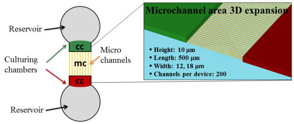
Background: Microglia possess an elevated grade of plasticity, undergoing several structural changes based on their location and state of activation. The first step towards the comprehension of microglia's biology and functional responses to an extremely mutable extracellular milieu, consists in discriminating the morphological features acquired by cells maintained in vitro under diverse environmental conditions. Previous work described neither primary microglia grown on artificially patterned environments which impose physical cues and constraints, nor long distance migration of microglia in vitro. To this aim, the present work exploits artificial bio-mimetic microstructured substrates with pillar-shaped or line-grating geometries fabricated on poly(dimethylsiloxane) by soft lithography, in addition to microfluidic devices, and highlights some morphological/functional characteristics of microglia which were underestimated or unknown so far.

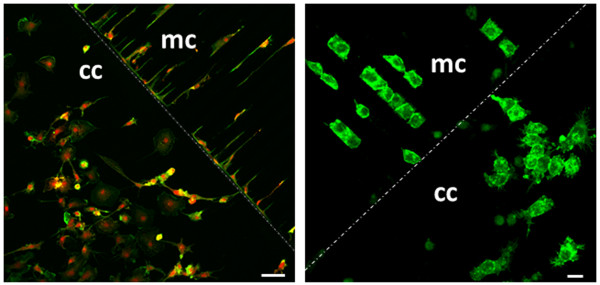
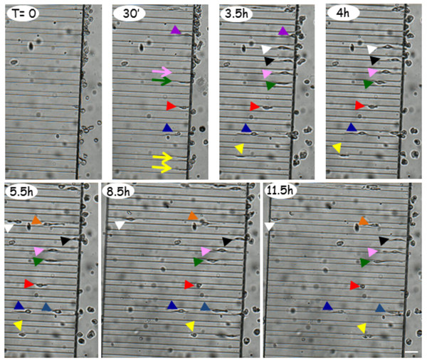
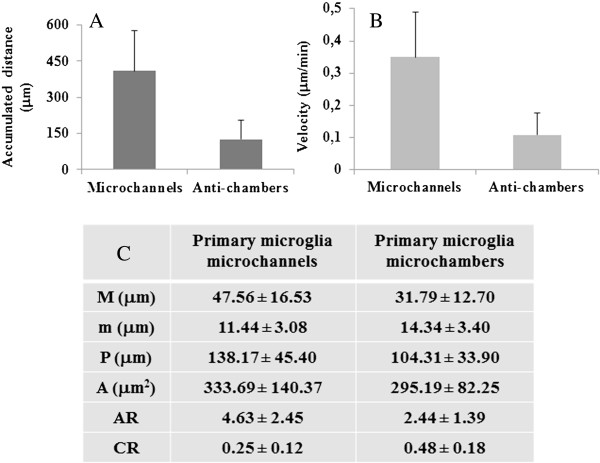
Results: We report that primary microglia selectively adapt to diverse microstructured substrates modifying accordingly their morphological features and behavior. On micropatterned pillar-shaped geometries, microglia appear multipolar, extend several protrusions in all directions and form distinct pseudopodia. On both micropatterned line-grating geometries and microfluidic channels, microglia extend the cytoplasm from a roundish to a stretched, flattened morphology and assume a filopodia-bearing bipolar structure. Finally, we show that in the absence of any applied chemical gradient, primary microglia spontaneously moves through microfluidic channels for a distance of up to 500 μm in approximately 12 hours, with an average speed of 0.66 μm/min.

Conclusions: We demonstrate an elevated grade of microglia plasticity in response to a mutable extracellular environment, thus making these cells an appealing population to be further exploited for lab on chip technologies. The development of microglia-based microstructured substrates opens the road to novel hybrid platforms for testing drugs for neuroinflammatory diseases.


Figures

Figure 1
Figure 1
Micropatterned structures serving for primary microglia culturing. Microtopographic silicon masters are realized to replica-mold substrates on PDMS with pillar-shaped and line-grating geometries (width: 1500 nm, pitch: 3 μm, height: 550 nm). A. Scanning electron microscope (Zeiss EVO) images of PDMS pillar- shaped substrates. B. Scanning electron microscope images of Si master with line-grating structures. C. PDMS line-gratings at high magnification.
Figure 2
Figure 2
Microglia adjust to micropatterned structures. A. Primary mouse microglia are cultured on plastic dishes and subjected to immunofluorescence and confocal analysis with phalloidin (green) plus P2Y12 receptors antiserum (red) and Hoechst (blue), scale bar = 50 μm. B-C. Primary microglia are cultured on pillar microstructures subjected to fluorescence and confocal analysis with phalloidin (green) plus Hoechst (blue), scale bar = 20 μm (B), or immunofluorescence with paxillin (red), scale bar = 10 μm (C). D. Primary mouse microglia are cultured on line-grating geometries and subjected to fluorescence and confocal analysis with phalloidin (green) plus Hoechst (blue), scale bar = 20 μm.
Figure 3
Figure 3
Morphometric analysis of microglia on micropatterned structures. Morphometric analysis of primary microglia cultured on pillar microstructures (A) and line-grating geometries (B) is performed using a customized Matlab code (C). Values are expressed as mean ± SD, with n = 14 cells on pillars or line-grating structures. A significative difference is obtained for all parameters by Mann–Whitney test (p < 0.0003). “M” indicates major axis and “m” minor axis of cell’s fitted ellipse (panels A, B), “A” cell area, “P” cell perimeter, “AR” aspect ratio, “CR” circularity ratio.
Figure 4
Figure 4
Microfluidic structure adopted for primary microglia culturing. Schematic representation of the microfluidic device used for the microglia motility experiments. Reservoirs, culturing chambers (cc) and microchannels (mc) areas are highlighted.
Figure 5
Figure 5
Morphological transition of microglia in microfluidic devices. Primary rat microglia (left panel) and N9 microglia cell line (right panel) are maintained in culture on microfluidic devices for 24 hours and subjected to immunofluorescence and confocal analysis with phalloidin (green) plus P2Y12 receptors antiserum (red) (left panel, scale bar = 50 μm) or fluorescence with phalloidin (right panel, scale bar = 20 μm). mc indicates the microchannels areas and cc shows the culturing chambers, as schematically depicted in Figure 4.
Figure 6
Figure 6
Migration of primary microglia in microfluidic devices occurs on homogeneous fibronectin coating. A. A homogeneous fibronectin coating is formed inside the microchannels. B. The profile of fluorescence intensity acquired with Zen software of Zeiss LSM 700 microscope provides values ranging from 3 to 55 fluorescence intensity arbitrary units. C. Microglial cells are cultured in microfluidic devices and subjected to immunofluorescence for IBA1 (red) and staining with Hoechst (blue). The total migration path is: length 500 μm (white dotted arrow), width 12 μm, height 10 μm, and the scale bar is 50 μm.
Figure 7
Figure 7
Primary microglia spontaneously migrate in microfluidic channels. After plating primary microglia in microfluidic devices for 30 min, time-lapse recording is performed every 30 min for 20 h. Arrows represent microglial processes and arrowheads indicate microglial cells. The different colors point to different forward- or backward-displacement of the cells inside the microchannels. The total migration path is: length 500 μm (white dotted arrow), width 12 μm, height 10 μm. The scale bar is 50 μm.
Figure 8
Figure 8
Morphometric analysis of primary microglia migrating into microfluidic channels. A. Mean accumulated distance of microglia somata over 20 h time-lapse recording. B. Mean velocity (μm/min) of microglia somata in microfluidic devices. C. Randomly selected cells moving in the microchannels (n = 79 cells) and culturing chamber (n = 120 cells) are manually tracked. Image stacks and time series are analyzed by Image J software. Data presented as mean ± SD. Statistical difference is obtained by Mann–Whitney test (p < 0.0003). “M” is major axis, “m” minor axis, “P” cell perimeter, “A” cell area, “AR” aspect ratio, “CR” circularity ratio.

Similar articles

Cited by

References

    1. del Rio-Hortega P. In: Cytology and Cellular Pathology of the Nervous System. Penfield W, editor. New York: Hoeber; 1932. Microglia; pp. 482–534.
    1. Nimmerjahn A, Kirchhoff F, Helmchen F. Resting microglial cells are highly dynamic surveillants of brain parenchyma in vivo. Science. 2005;308:1314–1318. doi: 10.1126/science.1110647. - DOI - PubMed
    1. Rezaie P, Male D. Colonisation of the developing human brain and spinal cord by microglia: a review. Microsc Res Tech. 1999;45:359–382. doi: 10.1002/(SICI)1097-0029(19990615)45:6<359::AID-JEMT4>3.0.CO;2-D. - DOI - PubMed
    1. Wake H, Moorhouse AJ, Nabekura J. Functions of microglia in the central nervous system–beyond the immune response. Neuron Glia Biol. 2011;7:47–53. doi: 10.1017/S1740925X12000063. - DOI - PubMed
    1. Ransohoff RM, Brown MA. Innate immunity in the central nervous system. J Clin Invest. 2012;122:1164–1171. doi: 10.1172/JCI58644. - DOI - PMC - PubMed

Publication types

LinkOut - more resources