

Anti-Müllerian hormone may regulate the number of calbindin-positive neurons in the sexually dimorphic nucleus of the preoptic area of male mice
- PMID: 24119315
- PMCID: PMC3852321
- DOI: 10.1186/2042-6410-4-18
Anti-Müllerian hormone may regulate the number of calbindin-positive neurons in the sexually dimorphic nucleus of the preoptic area of male mice
Abstract
Background: The male brain is putatively organised early in development by testosterone, with the sexually dimorphic nucleus of the medial preoptic area (SDN) a main exemplifier of this. However, pubescent neurogenesis occurs in the rat SDN, and the immature testes secrete anti-Müllerian hormone (AMH) as well as testosterone. We have therefore re-examined the development of the murine SDN to determine whether it is influenced by AMH and/or whether the number of calbindin-positive (calbindin+ve) neurons in it changes after pre-pubescent development.
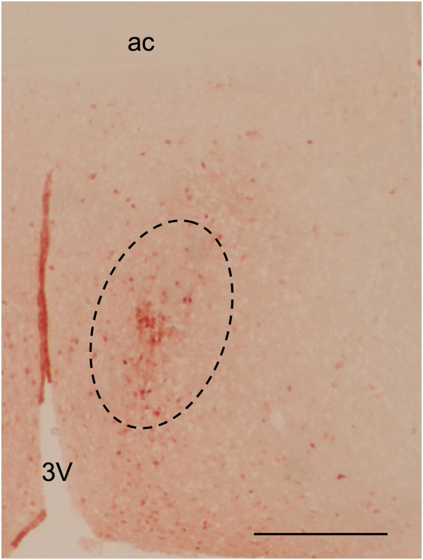
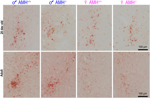
Methods: In mice, the SDN nucleus is defined by calbindin+ve neurons (CALB-SDN). The number and size of the neurons in the CALB-SDN of male and female AMH null mutant (Amh-/-) mice and their wild-type littermates (Amh+/+) were studied using stereological techniques. Groups of mice were examined immediately before the onset of puberty (20 days postnatal) and at adulthood (129-147 days old).
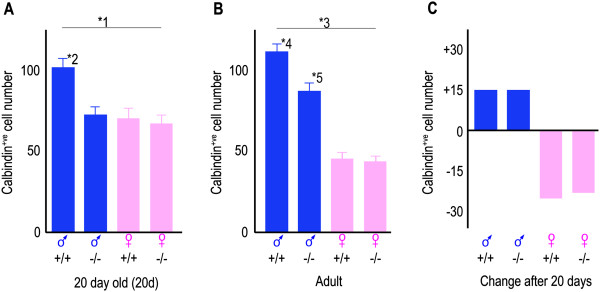
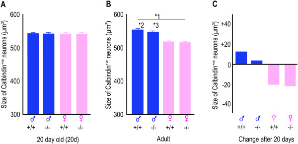
Results: The wild-type pre-pubertal male mice had 47% more calbindin+ve neurons in the CALB-SDN than their female wild-type littermates. This sex difference was entirely absent in Amh-/- mice. In adults, the extent of sexual dimorphism almost doubled due to a net reduction in the number and size of calbindin+ve neurons in females and a net increase in neuron number in males. These changes occurred to a similar extent in the Amh-/- and Amh+/+ mice. Consequently, the number of calbindin+ve neurons in Amh-/- adult male mice was intermediate between Amh+/+ males and Amh+/+ females. The sex difference in the size of the neurons was predominantly generated by a female-specific atrophy after 20 days, independent of AMH.
Conclusions: The establishment of dimorphic cell number in the CALB-SDN of mice is biphasic, with each phase being subject to different regulation. The second phase of dimorphism is not dependent on the first phase having occurred as it was present in the Amh-/- male mice that have female-like numbers of calbindin+ve neurons at 20 days. These observations extend emerging evidence that the organisation of highly dimorphic neuronal networks changes during puberty or afterwards. They also raise the possibility that cellular events attributed to the imprinting effects of testosterone are mediated by AMH.
Figures
Similar articles
-
Emergence of the sex difference in calbindin D-28K neurons and the role of calbindin D-28K in clustering neurons of the preoptic area in mice.Cell Tissue Res. 2025 Aug;401(2):117-128. doi: 10.1007/s00441-025-03981-3. Epub 2025 May 21. Cell Tissue Res. 2025. PMID: 40397161 Free PMC article.
-
Effects of blocking developmental cell death on sexually dimorphic calbindin cell groups in the preoptic area and bed nucleus of the stria terminalis.Biol Sex Differ. 2012 Feb 15;3:5. doi: 10.1186/2042-6410-3-5. Biol Sex Differ. 2012. PMID: 22336348 Free PMC article.
-
Calbindin-D28k immunoreactivity is a marker for a subdivision of the sexually dimorphic nucleus of the preoptic area of the rat: developmental profile and gonadal steroid modulation.J Neuroendocrinol. 2000 May;12(5):397-402. doi: 10.1046/j.1365-2826.2000.00474.x. J Neuroendocrinol. 2000. PMID: 10792577
-
Sex differences and the roles of sex steroids in apoptosis of sexually dimorphic nuclei of the preoptic area in postnatal rats.J Neuroendocrinol. 2009 Mar;21(4):370-6. doi: 10.1111/j.1365-2826.2009.01855.x. J Neuroendocrinol. 2009. PMID: 19226350 Review.
-
Development of the sexually dimorphic nucleus of the preoptic area and the influence of estrogen-like compounds.Neural Regen Res. 2013 Oct 15;8(29):2763-74. doi: 10.3969/j.issn.1673-5374.2013.29.008. Neural Regen Res. 2013. PMID: 25206587 Free PMC article. Review.
Cited by
-
Emergence of the sex difference in calbindin D-28K neurons and the role of calbindin D-28K in clustering neurons of the preoptic area in mice.Cell Tissue Res. 2025 Aug;401(2):117-128. doi: 10.1007/s00441-025-03981-3. Epub 2025 May 21. Cell Tissue Res. 2025. PMID: 40397161 Free PMC article.
-
Spermatogonial Type 3 Deiodinase Regulates Thyroid Hormone Target Genes in Developing Testicular Somatic Cells.Endocrinology. 2019 Dec 1;160(12):2929-2945. doi: 10.1210/en.2019-00259. Endocrinology. 2019. PMID: 31621880 Free PMC article.
-
Sexually Dimorphic Formation of the Preoptic Area and the Bed Nucleus of the Stria Terminalis by Neuroestrogens.Front Neurosci. 2020 Jul 29;14:797. doi: 10.3389/fnins.2020.00797. eCollection 2020. Front Neurosci. 2020. PMID: 32848568 Free PMC article. Review.
-
Anti-Müllerian hormone: a new actor of sexual dimorphism in pituitary gonadotrope activity before puberty.Sci Rep. 2016 Mar 31;6:23790. doi: 10.1038/srep23790. Sci Rep. 2016. PMID: 27030385 Free PMC article.
-
Novel role for anti-Müllerian hormone in the regulation of GnRH neuron excitability and hormone secretion.Nat Commun. 2016 Jan 12;7:10055. doi: 10.1038/ncomms10055. Nat Commun. 2016. PMID: 26753790 Free PMC article.
References
-
- Collaer ML, Hines M. Human behavioral sex differences: a role for gonadal hormones during early development? Psychol Bull. 1995;118:55–107. - PubMed
LinkOut - more resources
Full Text Sources
Other Literature Sources
Molecular Biology Databases
