Comparative Study
. 2013 Oct 10;52(1):124-34.
doi: 10.1016/j.molcel.2013.08.020.

Structure of the CRISPR interference complex CSM reveals key similarities with cascade

Affiliations
Comparative Study

Structure of the CRISPR interference complex CSM reveals key similarities with cascade

Christophe Rouillon et al. Mol Cell. .

Abstract

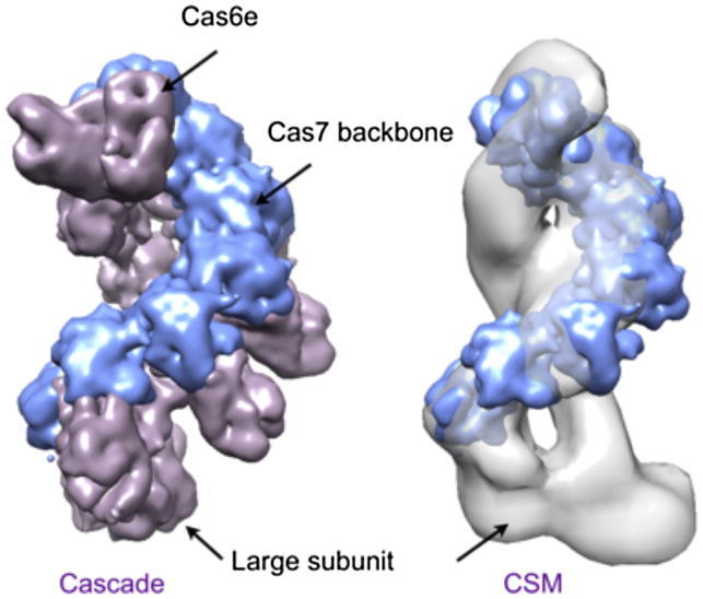
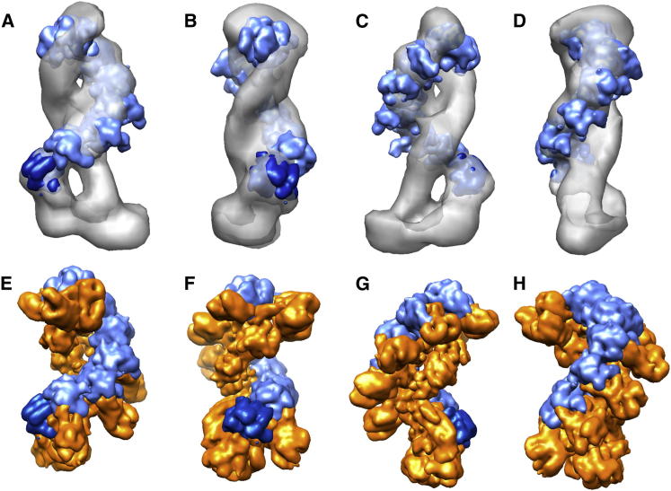
The Clustered Regularly Interspaced Palindromic Repeats (CRISPR) system is an adaptive immune system in prokaryotes. Interference complexes encoded by CRISPR-associated (cas) genes utilize small RNAs for homology-directed detection and subsequent degradation of invading genetic elements, and they have been classified into three main types (I-III). Type III complexes share the Cas10 subunit but are subclassifed as type IIIA (CSM) and type IIIB (CMR), depending on their specificity for DNA or RNA targets, respectively. The role of CSM in limiting the spread of conjugative plasmids in Staphylococcus epidermidis was first described in 2008. Here, we report a detailed investigation of the composition and structure of the CSM complex from the archaeon Sulfolobus solfataricus, using a combination of electron microscopy, mass spectrometry, and deep sequencing. This reveals a three-dimensional model for the CSM complex that includes a helical component strikingly reminiscent of the backbone structure of the type I (Cascade) family.


Figures

None
Graphical abstract
Figure 1
Figure 1
Purification and RNA Content of the CSM Complex from S. solfataricus (A) Gene organization of the csm locus in selected crenarchaeal species. The gene order is conserved and typically includes a gene encoding Cas6 for crRNA processing. Gene numbers are shown and are contiguous on the genome. Abbreviations are as follows: Sso, S. solfataricus; Sto, S. tokodaii; Sac, S. acidocaldarius; Sis, S. islandicus strain M.14.25; Msed, Metallosphaera sedula; Tpen, Thermofilum pendens. T. pendens Cas6 is present elsewhere on the genome. (B) Fractions of the CSM complex eluting from the final gel-filtration column during purification. All eight subunits can be visualized and detected by MS. (C) RNA purified from the purified CSM complex. A single discrete band around 50 nt was observed. (D) A linear coverage map for a series of eight spacers from the S. solfataricus P1 A locus is shown. 5′ ends were defined by the 8 nt 5′-handle derived from cleavage of the repeat by Cas6. (E) Linear coverage map for the entire CRISPR C locus from S. solfataricus P1, highlighting the variability in coverage.
Figure 2
Figure 2
3D-EM Reconstruction of the CSM Complex (A) Raw micrograph, with representative single particles in white circles. (B) Class averages and reprojections from the 3D reconstruction. (C) Surface representation of the full 3D CSM volume. (D) FSC plot.
Figure 3
Figure 3
MS Analysis of the CSM Complex Establishing Its Composition, Subunit Connectivity, and crRNA Binding (A) MS spectrum of the intact CSM reveals a well-resolved charge-state series at 8,500 m/z with a molecular mass of 427,789 Da, 122 kDa higher than the expected mass for a stoichiometric complex comprising eight subunits and one crRNA. (B) The 49+ charge state of the complex was selected and subjected to acceleration, and dissociation of subunits Sso1424, Sso1428, and Sso1426/7 was observed by tandem MS. (C) The molar ratio of Sso1426:Sso1428 was determined as 4:1 by relative quantification of tryptic peptides of Sso1426 and Sso1428 (GSVDLNYLR and FLDSLPISYSLNTR, respectively; see Table 1 and Table S3). Labeled peptides of the same sequences were synthesized and used as reference. (15N,13C)-labeled residues are colored red. (D–F) Disassembly of the CSM complex resulted in a series of subcomplexes (i–v) in solutions of decreasing pH: 3.9 (D), 3.5 (E), and 3.2 (F). (G) A complete CSM subunit interaction map was derived from MS data, including intact subcomplexes, crosslinking, and quantitative analysis (see also Figures S1–S3 and Tables S2–S6). The crRNA binds to subunits making up the major backbone and dissociates together with three copies of Sso1426 and Sso1432.
Figure 4
Figure 4
Ion Mobility Measurement of the CSM Complex and Its Subcomplexes (A) CCS values measured for the intact complex (purple circle), the 357 kDa (green diamond) and 216 kDa (orange square) subcomplexes, and the largest subunit Sso1428 (red triangle) are plotted against their masses. Three trendlines are shown for linear, linear dimer, or collapsed “globular” conformations (left to right) for complexes composed of monomers (25 kDa). Considerable deviation from all conformation is evident for the intact complex and the two subcomplexes. (B–D) Coarse-grain structural models, calculated for the intact complexes (D) and the 357 kDa (C) and 216 kDa (B) subcomplexes and fitted into the CSM EM map. Each subunit is represented by a sphere, sized proportionally to its mass, except that the largest Sso1428 is divided into two domains.
Figure 5
Figure 5
Fitting the Cascade Backbone in CSM and Comparison of the Two Structures (A–D) Orthogonal views of CSM (gray surface) with fitted Cas5 (dark blue) and six Cas7 proximal domains (light blue). (E–H) Orthogonal views of the Cascade complex from E. coli, where Cas5 and Cas7 proximal domains have been colored blue for direct comparison with CSM.

Comment in

References

    1. Alber F., Kim M.F., Sali A. Structural characterization of assemblies from overall shape and subcomplex compositions. Structure. 2005;13:435–445. - PubMed
    1. Albers S.V., Jonuscheit M., Dinkelaker S., Urich T., Kletzin A., Tampé R., Driessen A.J., Schleper C. Production of recombinant and tagged proteins in the hyperthermophilic archaeon Sulfolobus solfataricus. Appl. Environ. Microbiol. 2006;72:102–111. - PMC - PubMed
    1. Blankenberg D., Von Kuster G., Coraor N., Ananda G., Lazarus R., Mangan M., Nekrutenko A., Taylor J. Galaxy: a web-based genome analysis tool for experimentalists. Curr. Protoc. Mol. Biol. 2010;Chapter 19 Unit 19.10.1–21. - PMC - PubMed
    1. Botting C.H., Talbot P., Paytubi S., White M.F. Extensive lysine methylation in hyperthermophilic crenarchaea: potential implications for protein stability and recombinant enzymes. Archaea. 2010;2010:106341. - PMC - PubMed
    1. Esser D., Pham T.K., Reimann J., Albers S.V., Siebers B., Wright P.C. Change of carbon source causes dramatic effects in the phospho-proteome of the archaeon Sulfolobus solfataricus. J. Proteome Res. 2012;11:4823–4833. - PubMed

Publication types

MeSH terms