MRI findings of radiation-induced changes in the urethra and periurethral tissues after treatment for prostate cancer
- PMID: 24119430
- PMCID: PMC5714318
- DOI: 10.1016/j.ejrad.2013.09.011
MRI findings of radiation-induced changes in the urethra and periurethral tissues after treatment for prostate cancer
Abstract
Purpose: To assess radiotherapy (RT)-induced changes in the urethra and periurethral tissues after treatment for prostate cancer (PCa).
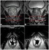
Methods and materials: This retrospective study included 108 men (median age, 64 years; range, 43-87 years) who received external-beam radiotherapy (EBRT) and/or brachytherapy for PCa and underwent endorectal-coil MRI of the prostate within 180 days before RT and a median of 20 months (range, 2-62 months) after RT. On all MRIs, two readers independently measured the urethral length (UL) and graded the margin definition (MD) of the urethral wall and the signal intensities (SIs) of the urethral wall and pelvic muscles on 4-point scales.
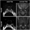
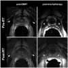
Results: The mean urethral length decreased significantly from pre- to post-RT MRI (from 15.2 to 12.6mm and from 14.4 to 12.9 mm for readers 1 and 2, respectively; both p-values <0.0001). Brachytherapy resulted in greater urethral shortening than EBRT. After RT, SI in the urethral wall increased in 57% (62/108) and 35% (38/108) of patients (readers 1 and 2, respectively). The frequency and magnitude of SI increase in pelvic muscles depended on muscle location. In the obturator internus muscle, SI increased more often after EBRT than after brachytherapy, while in the periurethral levator ani muscle SI increased more often after brachytherapy than after EBRT.
Conclusion: After RT for PCa, MRI shows urethral shortening and increased SI of the urethral wall and pelvic muscles in substantial percentages of patients.
Keywords: MRI; Prostate cancer, Urethra; Radiation therapy.
Copyright © 2013 Elsevier Ireland Ltd. All rights reserved.
Conflict of interest statement
Conflict of interest: none
Figures


References
-
- Zelefsky MJ, Wallner KE, Ling CC, et al. Comparison of the 5-year outcome and morbidity of three-dimensional conformal radiotherapy versus transperineal permanent iodine-125 implantation for early-stage prostatic cancer. Journal of clinical oncology: official journal of the American Society of Clinical Oncology. 1999;17(2):517–22. - PubMed
-
- Kupelian PA, Elshaikh M, Reddy CA, Zippe C, Klein EA. Comparison of the efficacy of local therapies for localized prostate cancer in the prostate-specific antigen era: a large single-institution experience with radical prostatectomy and external-beam radiotherapy. Journal of clinical oncology: official journal of the American Society of Clinical Oncology. 2002;20(16):3376–85. - PubMed
-
- Aluwini S, van Rooij PH, Kirkels WJ, et al. High-dose-rate brachytherapy and external-beam radiotherapy for hormone-naive low- and intermediate-risk prostate cancer: a 7-year experience. International journal of radiation oncology, biology, physics. 2012;83(5):1480–5. - PubMed
-
- Grimm P, Billiet I, Bostwick D, et al. Comparative analysis of prostate-specific antigen free survival outcomes for patients with low, intermediate and high risk prostate cancer treatment by radical therapy. Results from the Prostate Cancer Results Study Group. BJU international. 2012;109(Suppl 1):22–9. - PubMed
Publication types
MeSH terms
Grants and funding
LinkOut - more resources
Full Text Sources
Other Literature Sources
Medical

