Review
. 2014 Jan;24(1):61-72.
doi: 10.1016/j.tcb.2013.09.005. Epub 2013 Oct 24.

Immune synapse: conductor of orchestrated organelle movement

Affiliations
Review

Immune synapse: conductor of orchestrated organelle movement

Noa Beatriz Martín-Cófreces et al. Trends Cell Biol. 2014 Jan.

Abstract

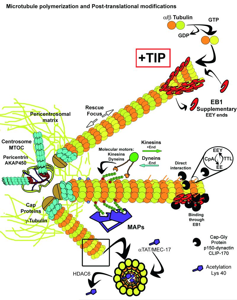
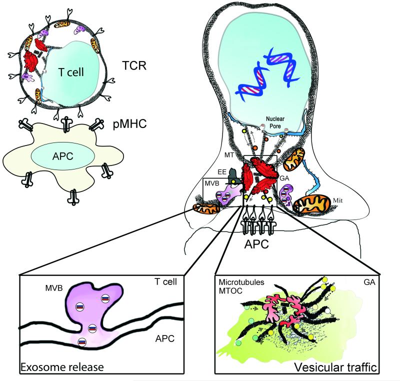
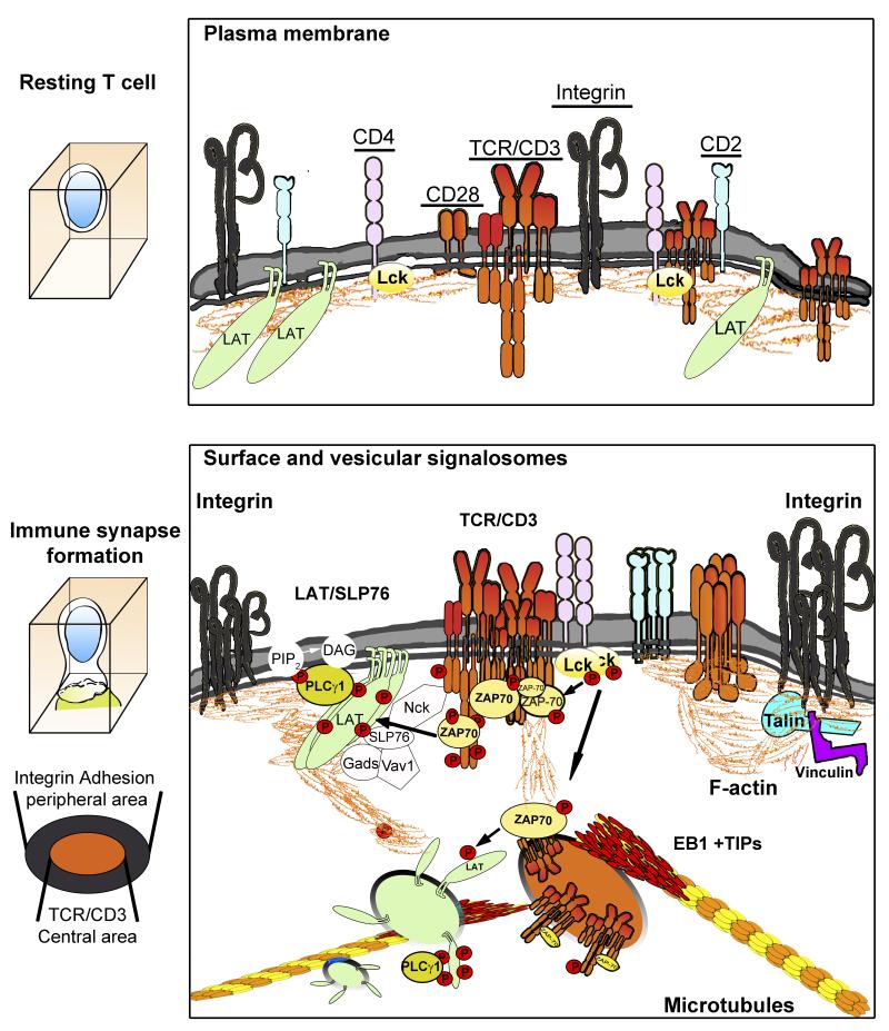
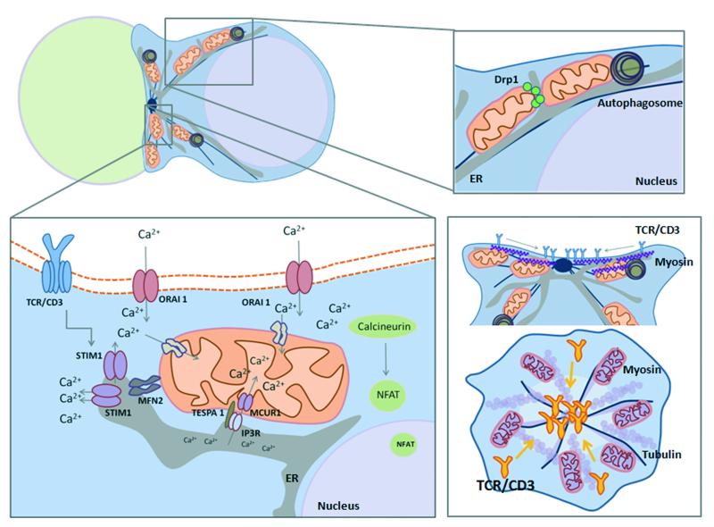
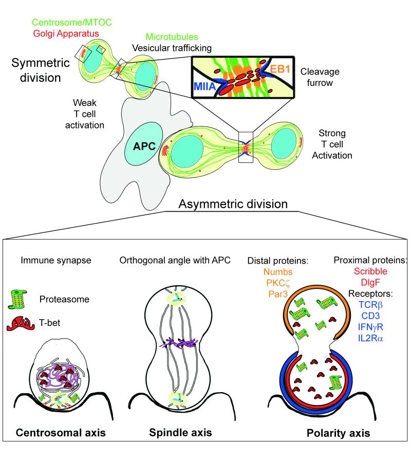
To ensure proper cell function, intracellular organelles are not randomly distributed within the cell, but polarized and highly constrained by the cytoskeleton and associated adaptor proteins. This relationship between distribution and function was originally found in neurons and epithelial cells; however, recent evidence suggests that it is a general phenomenon occurring in many highly specialized cells including T lymphocytes. Recent studies reveal that the orchestrated redistribution of organelles is dependent on antigen-specific activation of and immune synapse (IS) formation by T cells. This review highlights the functional implications of organelle polarization in early T cell activation and examines recent findings on how the IS sets the rhythm of organelle motion and the spread of the activation signal to the nucleus.

Keywords: immune synapse; microcluster; microtubule; mitochondria; signalosome; vesicle.


Figures

Figure I
Figure I
The microtubule network emerges from the centrosome and the pericentrosomal matrix allows the binding of γ-Tubulin Ring Complexes (γTuRC), formed by γ-Tubulin and capping proteins. In association with microtubule plus-end tracking proteins (+TIPS), the γTuRCs provide stable plus-ends that allow microtubules to grow. One such +TIPS, end-binding 1 rotein (EB1), facilitates the incorporation of α/β-Tubulin heterodimers into a sheet formed by the tubulin protofilaments, upon GDP interchange by GTP in the β-Tubulin subunit. EB proteins and proteins containing the TOG domain (XMAP125, CLASP) help fold the sheet into a tubule. The GTP then hydrolyzes into GDP and facilitates the catastrophe events that depolymerize the microtubules. However, “GTP seeds” may remain to serve as a rescue focus in preexisting microtubules.
Figure 1
Figure 1. Polarization of organelles at the immune synapse
The IS forms rapidly upon focal TCR activation. The TCR accumulates at the interface of the T cell and the stimulating surface; an antigen-presenting cell (APC) or a surface coated with stimulating antibodies or recombinant proteins, such as the MHC complex. The centrosome localizes to the IS and brings the associated Golgi Apparatus (GA). The early endosome compartment (EE) regulates recycling of surface receptors. The mitochondrial network (Mit), in association with the endoplasmic reticulum, dynamically localizes to the IS and provides a controlled calcium flux and a focused supply of ATP for energy provision. Nuclear activating molecules enter the nucleus via nuclear pores. They can be transported in vesicles or through direct interaction with molecular motors such as Dynein. Left inset: Multivesicular bodies (MVB) localize to the IS upon centrosome relocalization and have several functions, including the recycling of proteins and the formation of exosomes. MVBs serve as exocytosis compartments at the IS. Right inset: vesicular traffic at the IS. Vesicles coming from the IS are directed to recycling organelles, such as endosomes. While a number of vesicles emerging from the trans-Golgi network facilitate polarized secretion.
Figure 2
Figure 2. Crosstalk between signals and organelles of resting and activated T cells
In resting T cells, signaling receptors are evenly distributed throughout the plasma membrane. After the TCR engages an antigen-presenting cell, signaling receptors and molecules form microclusters, composed of different signalosomes, to propagate the signal. Below the plasma membrane, microtubule-dependent, subcortical vesicular trafficking helps to spread signals from the TCR toward the nucleus. The interchange of molecules between CD3, LAT and SLP76 clusters and CD3- and LAT-bearing vesicles can amplify these signaling pathways. The interaction of CD3 and LAT-containing vesicles is another mechanism for signal spread at the IS.
Figure 3
Figure 3. Mitochondrial polarization: fueling the immune synapse
Mitochondrial fission factor Drp1 localizes mitochondria towards the nascent IS. Increased ATP levels at the IS are needed for energy-consuming signalling and for the acto-myosin centripetal flux of TCR to the central area. The tethering of mitochondria with the endoplasmic reticulum (ER) is relevant for sustained Ca2+ influx upon TCR activation. Mitochondria avoid premature Ca2+-dependent inactivation of CRAC/ORAI1 channels and calcium flux across plasma membrane. Mitofusin-2-dependent mitochondria–ER tethering modulates trafficking of STIM1 to activate ORAI1. Additionally, Junctate has been proposed as an alternate Ca2+-sensing ER protein that regulates recruitment of stromal interaction molecule 1 (STIM1) [94]. Mitochondrial-Associated ER Membranes (MAMs) define the position of mitochondrial division sites where ER tubules contact mitochondria and mediate constriction before Drp1 recruitment [95]. MAMs regulate autophagosome formation through mitochondrial calcium uptake, which regulates cell bioenergetics and inhibits constitutive autophagy [72]. Tespa1 links IP3R-mediated Ca2+ release and mitochondrial Ca2+ uptake in the MAM compartment [96]. MCUR1 functions as a uniporter channel essential for calcium uptake in mitochondria [97]. TCR stimulation promotes autophagy and formation of autophagosomes. Autophagy regulates T cell homeostasis by promoting T lymphocyte survival and proliferation by orchestrating mitochondrial clearance in T cells. ER compartment is increased in autophagy-deficient T lymphocytes, which display a defective influx of calcium from expanded ER stores [98]. Impairing Drp1 function decreases the centripetal flux of the TCR to the central area through impairment of actomyosin contraction and maintains TCR activity, and promotes proximal TCR signalling and IL-2 production. Mitochondria play a dual role at the IS by regulating the strength of T cell activation and the termination of the IS signalling.
Figure 4
Figure 4. Asymmetric cell division in T cells
T cells can divide asymmetrically while in contact with an APC in vitro. Sustained synapses promoted by strong antigenic stimulation are critical for T cell activation and provide a cue for the establishment and maintenance of polarity throughout cell division. The cell asymmetry of proteins and organelles obtained through the challenge of the IS may be useful to conform an axis of polarity during cell division. The initial localization of the centrosome may be a marker to organize the spindle axis once the centrioles have duplicated. The polarity axis conformed by the IS can be reinforced by vesicular traffic from the Golgi towards the cleavage furrow during cell division. This traffic can promote the accumulation of proteins in one of the cells. EB1 parallels its own localization at the IS in the cleavage furrow. T cell receptors are unequally divided among daughter cells. CD4 naïve T cells promote TCRβ, IFNγR and IL2Rα polarization toward the IS and subsequently, the proximal cell [99]. CD3 and IL2Rα are similarly recruited in CD8 memory cells [100]. The polarity proteins known as partitioning defective (Par) proteins consist of the Par3-Par6-atypical PKC (aPKC) and Discs large (Dlg)-Scribble-Lethal giant larvae (Lgl) polarity complexes and are differentially distributed during asymmetric cell division, as is Numb, an endocytic adaptor that can also help to regulate polarity [80]. Indeed, several transcriptional regulators undergo asymmetric segregation in lymphocytes. T-bet, which drives effector differentiation in CD8+ T cells and the Th1 fate in CD4+ T cells is unequally inherited during division of both types of lymphocytes [101]. The asymmetric segregation of the proteasome determines the preferential degradation of T-bet, yielding one daughter cell that inherits more T-bet. These results suggest a mechanism during IS formation where a cell may unequally localize cellular activities during division, thereby imparting disparity in the abundance of cell fate regulators in the daughter cells.

References

    1. Lasserre R, Alcover A. Cytoskeletal cross-talk in the control of T cell antigen receptor signaling. FEBS Lett. 2010;584:4845–4850. - PubMed
    1. Martin-Cofreces NB, et al. Tubulin and actin interplay at the T cell and antigen-presenting cell interface. Front Immunol. 2011;2:24. - PMC - PubMed
    1. Shaw AS, Filbert EL. Scaffold proteins and immune-cell signalling. Nat Rev Immunol. 2009;9:47–56. - PubMed
    1. Yanez-Mo M, et al. Tetraspanin-enriched microdomains: a functional unit in cell plasma membranes. Trends Cell Biol. 2009;19:434–446. - PubMed
    1. Baixauli F, et al. The mitochondrial fission factor dynamin-related protein 1 modulates T-cell receptor signalling at the immune synapse. EMBO J. 2011;30:1238–1250. - PMC - PubMed

Publication types

LinkOut - more resources