. 2013 Nov 7;93(5):906-14.
doi: 10.1016/j.ajhg.2013.09.011. Epub 2013 Oct 10.

Macrocytic anemia and mitochondriopathy resulting from a defect in sideroflexin 4

Affiliations

Macrocytic anemia and mitochondriopathy resulting from a defect in sideroflexin 4

Gordon J Hildick-Smith et al. Am J Hum Genet. .

Abstract

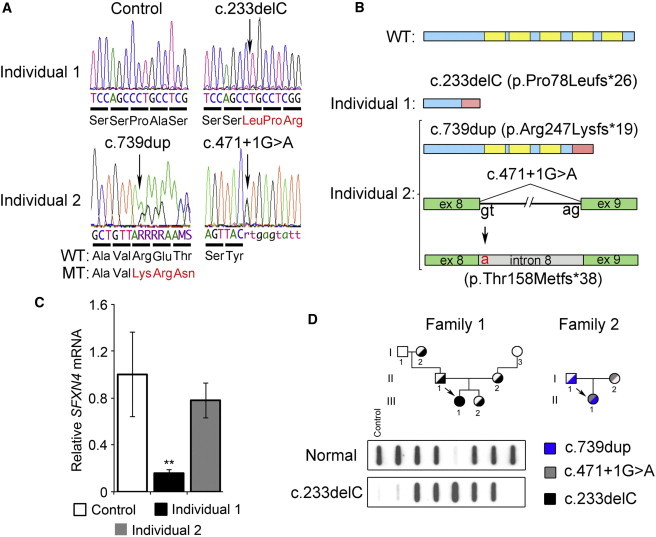
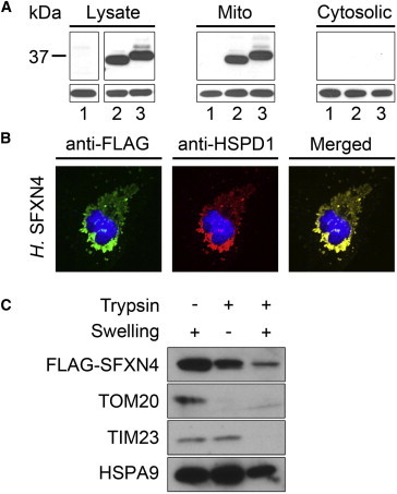
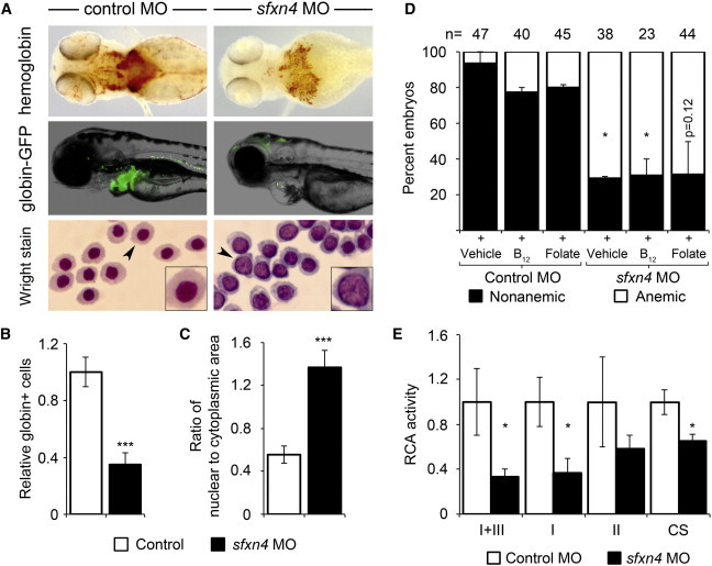
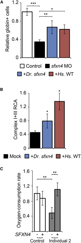
We used exome sequencing to identify mutations in sideroflexin 4 (SFXN4) in two children with mitochondrial disease (the more severe case also presented with macrocytic anemia). SFXN4 is an uncharacterized mitochondrial protein that localizes to the mitochondrial inner membrane. sfxn4 knockdown in zebrafish recapitulated the mitochondrial respiratory defect observed in both individuals and the macrocytic anemia with megaloblastic features of the more severe case. In vitro and in vivo complementation studies with fibroblasts from the affected individuals and zebrafish demonstrated the requirement of SFXN4 for mitochondrial respiratory homeostasis and erythropoiesis. Our findings establish mutations in SFXN4 as a cause of mitochondriopathy and macrocytic anemia.


Figures

Figure 1
Figure 1
Individual 1 Has Macrocytic Anemia, and Both Individuals Present with Mitochondriopathy (A) The RCA defect was seen in primary fibroblasts from both individuals, but not in control fibroblasts. RCA activity was assayed as described. Error bars represent the SEM. (B) Immunoblot analyses of mitochondrial-respiratory-complex proteins from control fibroblasts (C1, C2, and C3) and fibroblasts from the two affected individuals (I1 and I2) are displayed. (C) Densitometric quantification of the respiratory-complex proteins, shown in (B), demonstrates intact expression of the respiratory-chain components. Error bars represent the SEM. (D) Peripheral-blood smears from individual 1 reveal erythroid macrocytosis (left, arrow heads) and hypersegmented neutrophils (right, arrow).
Figure 2
Figure 2
Next-Generation Sequencing Identifies Mutations in SFXN4 (A) Upper panel: sequencing revealed a single-nucleotide deletion (c.233delC) in the SFXN4 cDNA from individual 1 (right), but not in the control (left). Lower panel: sequencing of individual 2 revealed two mutations, c.739dup (left) and c.471+1G>A (right). The normal and aberrant amino acid sequences are displayed below each chromatogram. (B) A schematic representation of the normal SFXN4 is shown with the predicted multispanning transmembrane domains (top, highlighted in yellow). The c.233delC mutation in individual 1 results in a frameshift (highlighted in pink) and an eventual nonsense substitution at amino acid residue 102 (p.Pro78Leufs26). In individual 2, the c.739dup mutation also causes a frameshift (pink) and premature termination at amino acid 266 (p.Arg247Lysfs19), whereas the c.471+1G>A mutation affects the splice donor site at intron 8, causes its retention in the misspliced mRNA, and thus results in the insertion of an additional 38 amino acids after Tyr157 (p.Thr158Metfs38). (C) qRT-PCR analysis from control and affected fibroblasts revealed decreased steady-state SFXN4 mRNA when normalized to glyceraldehyde-3-phosphate dehydrogenase (GAPDH), correlating the disease severity with the residual SFXN4 mRNA level; ∗∗p < 0.0005. qRT-PCR was performed with TaqMan probes for SFXN4 and GAPDH (Applied Biosystems, Life Technologies). SFXN4 expression was analyzed with the standard curve method, and GAPDH was used as a normalization control. Error bars represent the SEM. (D) Family 1: Mendelian recessive-inheritance segregation was demonstrated in the family of individual 1 (arrow, III-1) by allele-specific oligonucleotide hybridization with the normal (top) or mutant c.233delC (bottom) 32P-labeled probes. The control is a GM00536A healthy male. Family 2: the compound heterozygosity of individual 2 (arrow, II-2) was established with Sanger dideoxy sequencing.
Figure 3
Figure 3
SFXN4 Localizes to the Inner Mitochondrial Membrane (A) With the use of Lipofectamine 2000 (Invitrogen) and immunoblot analysis with FLAG antibody (Sigma) (lane 1, mock transfection; lane 2, zebrafish sfxn4; lane 3, human SFXN4), FLAG-tagged SFXN4 from human and zebrafish was found to localize to the mitochondria in transiently transfected Cos7 cells. Lysate refers to total cellular lysate, and mito (mitochondrial) and cytosolic refer to the subcellular fractions. HSPD1 (total and mitochondrial; Santa Cruz) and GAPDH (cytosolic; Santa Cruz) antibodies were used as loading controls. Cells were prepared for immunoblotting as previously described. FLAG antibodies were obtained from Sigma. Goat anti-mouse IgG-HRP was used as a secondary antibody. Proteins were visualized with the SuperSignal West Pico Substrate chemiluminescent (Pierce). (B) Confocal immunofluorescence microscopy confirmed the colocalization of FLAG-SFXN4 (FITC, green) with HSPD1 (Texas Red) (Pearson’s correlation coefficient, 0.74), a mitochondria resident protein (yellow, merged panel). Nuclei were stained with DAPI (blue). The correlation between the signal from the FLAG-tagged SFXN4 and mitochondrial marker HSPD1 was calculated with software developed by Tony Collins, Wayne Rasband, and Kevin Baler. The Pearson’s coefficient was compared against 74 randomized iterations of the HSPD1 images via the Fay method and was statistically significant (p < 0.05). FITC and Texas-Red-conjugated secondary antibodies were obtained from Santa Cruz. (C) FLAG-SFXN4 localized to the inner mitochondrial membrane in transfected HeLa cells after trypsin digestion (lane 2). TIM23, an inner-membrane protein, was protected from trypsin digestion, whereas TOM20, an outer-membrane protein, was degraded (lane 2). Rupturing of the mitochondrial inner membrane by hypotonic treatment showed that FLAG-SFXN4 and TIM23 were sensitive to proteolysis, whereas HSPA9, a matrix protein was protected (lane 3). The residual protease-resistant FLAG-SFXN4 reflects mitochondria that are resistant to osmotic shock. Proteins were detected with FLAG (Gilbertsville), TOM20 (Santa Cruz), TIM23 (BD Biosciences), and HSPA9 (Santa Cruz) antibodies.
Figure 4
Figure 4
sfxn4 Knockdown in Zebrafish Recapitulates the Defects in Erythropoiesis and Mitochondrial Respiration Seen in Individual 1 (A) Phenotypic characterization of control and zebrafish morphants for sfxn4. Zebrafish sfxn4 morphants showed a defect in hemoglobinization (brown color) when stained with o-dianisidine (upper). The anemia in sfxn4 morphants was evident by the reduction of GFP+ erythroid cells in the Tg(globin-LCR:eGFP) transgenic zebrafish reporter line (middle). Cytospin analyses of flow-sorted erythroid cells from the control (left) and sfxn4 morphant (right) revealed large nuclei with noncondensed chromatin in the latter (lower). Enlarged magnifications of individual cells (arrow) are shown in the insets. (B) Flow cytometry of the Tg(globin-LCR:eGFP) transgenic line quantified the anemia in sfxn4 morphants with a splice-blocking MO (∗∗∗p < 0.0005). Cells were collected from 20–100 control or MO-injected embryos, disaggregated and passed sequentially through 70 and 40 μm cell strainers, washed in Hank’s Balanced Salt Solution (HBSS) (Sigma), and pelleted by low-speed centrifugation. The cells were resuspended in a final buffer containing HBSS. Cells were sorted in a BD Biosciences FACSVantage SE machine as described. Error bars represent the SEM. (C) Enumeration of the ratio of nuclear area to cytoplasmic area showed a large increase in nuclear size relative to residual cytoplasmic area (∗∗∗p < 0.0005; n > 250 cells were analyzed for each condition). Erythrocytes were sorted from embryos as previously described and stained with Wright-Giemsa dye in a clinical hematology laboratory (Dana-Farber Cancer Institute, Boston). Cells were individually given a chromatic threshold for designating nuclear and cytoplasmic regions. The nuclear and cytoplasmic areas were measured in Image J (National Institutes of Health) with investigator-coded software. Analysis was confirmed by manual inspection of all samples, and cellular components given improper thresholds were excluded from the analysis. Nuclear and cytoplasmic areas were averaged from a minimum of 250 cells (total) from four independent samples and divided for obtaining the ratio of nuclear area to cytoplasmic area. Statistical significance was established with a one-tailed Student’s t test for paired samples. Error bars represent the SEM. (D) Neither vitamin B12 (1.0 mM) nor folate (0.01 mM) chemically complemented the anemia in sfxn4 morphant zebrafish raised in vehicle media (standard balanced salt solution). The vehicle- and vitamin B12-treated sfxn4 morphants were significantly more anemic than the control morphant samples exposed to either vehicle or vitamin B12 (p < 0.05). Similarly, compared to morphant controls exposed to folate, folate-treated sfxn4 morphants were anemic (p = 0.12). Doses were selected on the basis of established literature and adjusted for preventing developmental and chemical toxicity. At 96 hr postfertilization, embryos were stained for hemoglobinized cells with o-dianisidine (Sigma) as described. Error bars represent the SEM. (E) Zebrafish sfxn4 morphants, but not embryos injected with a standard control MO, exhibited global mitochondrial-respiratory-chain defects (complexes I and III, complex I, complex II, citrate synthase3,27) (p < 0.05). Error bars represent the SEM.
Figure 5
Figure 5
SFXN4 Is Functionally Conserved across Vertebrate Species in Both Erythropoiesis and Mitochondrial Respiration (A) Normal SFXN4 cRNA from either zebrafish or human partially complemented the anemia in sfxn4 morphant embryos (∗∗p < 0.005, p < 0.05). Zebrafish and human FLAG-SFXN4 cDNA constructs in pXT7 were used for generating 5′ capped mRNA with the use of the mMessage mMachine T7 Kit (Ambion). The generated mRNA was mixed with MO at an equimolar concentration to the MO injection and injected at the 1-cell stage. The translational expression of the transgenic mRNA was confirmed by immunoblot analysis with FLAG antisera. (B) Normal SFXN4 cDNA from zebrafish and human complemented the RCA defect in primary fibroblasts of individual 1 (p < 0.05). RCA for complexes I and III were compared to a mock-transfected (empty-vector) sample. Transfections were carried out with 5 μg of human or zebrafish SFXN4 cDNA or empty vector pCS2+ with the use of Lipofectamine 2000 (Invitrogen). (C) Lentiviral transduction of individuals 2’s fibroblasts (performed as described32) with SFXN4 cDNA (DNASU Plasmid Repository, clone ID HsCD00352377) cloned into the pLenti6/3/V5-TOPO vector system (Invitrogen) complemented the complex I respiratory deficiency. The oxygen-consumption rate was measured as previously described after nontransduction (−) or SFXN4 stable transduction with lentivirus (+) on fibroblasts from the control and individual 2 (∗∗p < 0.01). Error bars represent the SEM.

References

    1. Vafai S.B., Mootha V.K. Mitochondrial disorders as windows into an ancient organelle. Nature. 2012;491:374–383. - PubMed
    1. Aslinia F., Mazza J.J., Yale S.H. Megaloblastic anemia and other causes of macrocytosis. Clin. Med. Res. 2006;4:236–241. - PMC - PubMed
    1. DiMauro S., Servidei S., Zeviani M., DiRocco M., DeVivo D.C., DiDonato S., Uziel G., Berry K., Hoganson G., Johnsen S.D. Cytochrome c oxidase deficiency in Leigh syndrome. Ann. Neurol. 1987;22:498–506. - PubMed
    1. Yu H.C., Sloan J.L., Scharer G., Brebner A., Quintana A.M., Achilly N.P., Manoli I., Coughlin C.R., 2nd, Geiger E.A., Schneck U. An X-linked cobalamin disorder caused by mutations in transcriptional coregulator HCFC1. Am. J. Hum. Genet. 2013;93:506–514. - PMC - PubMed
    1. Gherasim C., Lofgren M., Banerjee R. Navigating the B(12) road: assimilation, delivery, and disorders of cobalamin. J. Biol. Chem. 2013;288:13186–13193. - PMC - PubMed

Publication types