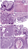
Benign papillomas without atypia diagnosed on core needle biopsy: experience from a single institution and proposed criteria for excision
- PMID: 24119786
- PMCID: PMC4605914
- DOI: 10.1016/j.clbc.2013.08.007
Benign papillomas without atypia diagnosed on core needle biopsy: experience from a single institution and proposed criteria for excision
Abstract
Background: The management of benign papilloma (BP) without atypia identified on breast core needle biopsy (CNB) is controversial. In this study, we determined the upgrade rate to malignancy for BPs without atypia diagnosed on CNB and whether there are factors associated with upgrade.
Methods: Through our pathology database search, we studied 80 BPs without atypia identified on CNB from 80 patients from 1997 to 2010, including 30 lesions that had undergone excision and 50 lesions that had undergone ≥ 2 years of radiologic follow-up. Associations between surgery or upgrade to malignancy and clinical, radiologic, and pathologic features were analyzed.
Results: Mass lesions, lesions sampled by ultrasound-guided CNB, and palpable lesions were associated with surgical excision. All 3 upgraded cases were mass lesions sampled by ultrasound-guided CNB. None of the lesions with radiologic follow-up only were upgraded to malignancy. The overall upgrade rate was 3.8%. None of the clinical, radiologic, or histologic features were predictive of upgrade.
Conclusion: Because the majority of patients can be safely managed with radiologic surveillance, a selective approach for surgical excision is recommended. Our proposed criteria for excision include pathologic/radiologic discordance or sampling by ultrasound-guided CNB without vacuum assistance when the patient is symptomatic or lesion size is ≥ 1.5 cm.
Keywords: Breast; Core needle biopsy; Criteria; Papilloma; Upgrade.
Copyright © 2013 Elsevier Inc. All rights reserved.
Conflict of interest statement
The authors have stated that they have no conflicts of interest.
Figures

Similar articles
-
Analysis of 612 Benign Papillomas Diagnosed at Core Biopsy: Rate of Upgrade to Malignancy, Factors Associated With Upgrade, and a Proposal for Selective Surgical Excision.AJR Am J Roentgenol. 2021 Dec;217(6):1299-1311. doi: 10.2214/AJR.21.25832. Epub 2021 May 19. AJR Am J Roentgenol. 2021. PMID: 34008998
-
Benign breast papillomas without atypia diagnosed with core needle biopsy: Outcome of surgical excision and imaging follow-up.Eur J Radiol. 2020 Oct;131:109237. doi: 10.1016/j.ejrad.2020.109237. Epub 2020 Aug 28. Eur J Radiol. 2020. PMID: 32905954
-
Asymptomatic Benign Papilloma Without Atypia Diagnosed at Ultrasonography-Guided 14-Gauge Core Needle Biopsy: Which Subgroup can be Managed by Observation?Ann Surg Oncol. 2016 Jun;23(6):1860-6. doi: 10.1245/s10434-016-5144-0. Epub 2016 Feb 26. Ann Surg Oncol. 2016. PMID: 26920388
-
Radial Sclerosing Lesion (Radial Scar): Radiologic-Pathologic Correlation.J Breast Imaging. 2024 Nov 5;6(6):646-657. doi: 10.1093/jbi/wbae046. J Breast Imaging. 2024. PMID: 39209731 Review.
-
Risk-Associated Lesions of the Breast in Core Needle Biopsies: Current Approaches to Radiological-Pathological Correlation.Surg Pathol Clin. 2022 Mar;15(1):147-157. doi: 10.1016/j.path.2021.11.010. Surg Pathol Clin. 2022. PMID: 35236630 Review.
Cited by
-
How Does Diagnostic Accuracy Evolve with Increased Breast MRI Experience?Tomography. 2023 Nov 6;9(6):2067-2078. doi: 10.3390/tomography9060162. Tomography. 2023. PMID: 37987348 Free PMC article.
-
Are we overtreating intraductal papillomas?J Surg Res. 2018 Nov;231:387-394. doi: 10.1016/j.jss.2018.06.008. Epub 2018 Jun 29. J Surg Res. 2018. PMID: 30278958 Free PMC article.
-
How Do We Approach Benign Proliferative Lesions?Curr Oncol Rep. 2018 Mar 23;20(4):34. doi: 10.1007/s11912-018-0682-1. Curr Oncol Rep. 2018. PMID: 29572753 Review.
-
Predictive Factors for Upgrading Patients with Benign Breast Papillary Lesions Using a Core Needle Biopsy.J Breast Cancer. 2016 Dec;19(4):410-416. doi: 10.4048/jbc.2016.19.4.410. Epub 2016 Dec 23. J Breast Cancer. 2016. PMID: 28053629 Free PMC article.
-
The genetic architecture of breast papillary lesions as a predictor of progression to carcinoma.NPJ Breast Cancer. 2020 Mar 12;6:9. doi: 10.1038/s41523-020-0150-6. eCollection 2020. NPJ Breast Cancer. 2020. PMID: 32195332 Free PMC article.
References
-
- Liberman L, Bracero N, Vuolo MA, et al. Percutaneous large-core biopsy of papillary breast lesions. AJR Am J Roentgenol. 1999;172:331–7. - PubMed
-
- Mercado CL, Hamele-Bena D, Singer C, et al. Papillary lesions of the breast: evaluation with stereotactic directional vacuum-assisted biopsy. Radiology. 2001;221:650–5. - PubMed
-
- Kim MJ, Kim SI, Youk JH, et al. The diagnosis of non-malignant papillary lesions of the breast: comparison of ultrasound-guided automated gun biopsy and vacuum-assisted removal. Clin Radiol. 2011;66:530–5. - PubMed
-
- Lakhani SR, Ellis IO, Schnitt SJ, et al. WHO Classification of Tumours of the Breast. 4. Lyon, France: International Agency for Research on Cancer (IARC); 2012. World Health Organization Classification of Tumours.
-
- Lam WW, Chu WC, Tang AP, et al. Role of radiologic features in the management of papillary lesions of the breast. AJR Am J Roentgenol. 2006;186:1322–7. - PubMed
Publication types
MeSH terms
Grants and funding
LinkOut - more resources
Full Text Sources
Other Literature Sources
Medical

