. 2014 Jul 16;32(33):4234-42.
doi: 10.1016/j.vaccine.2013.09.061. Epub 2013 Oct 10.

Protein coated microcrystals formulated with model antigens and modified with calcium phosphate exhibit enhanced phagocytosis and immunogenicity

Affiliations

Protein coated microcrystals formulated with model antigens and modified with calcium phosphate exhibit enhanced phagocytosis and immunogenicity

Sarah Jones et al. Vaccine. .

Abstract

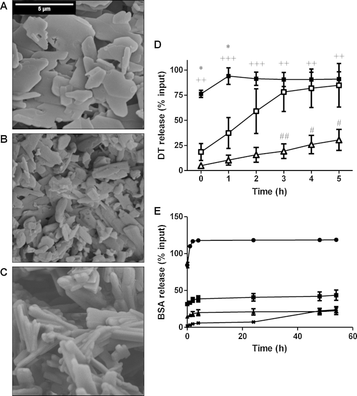
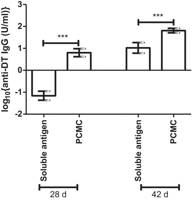
Protein-coated microcrystals (PCMCs) were investigated as potential vaccine formulations for a range of model antigens. Presentation of antigens as PCMCs increased the antigen-specific IgG responses for all antigens tested, compared to soluble antigens. When compared to conventional aluminium-adjuvanted formulations, PCMCs modified with calcium phosphate (CaP) showed enhanced antigen-specific IgG responses and a decreased antigen-specific IgG1:IgG2a ratio, indicating the induction of a more balanced Th1/Th2 response. The rate of antigen release from CaP PCMCs, in vitro, decreased strongly with increasing CaP loading but their immunogenicity in vivo was not significantly different, suggesting the adjuvanticity was not due to a depot effect. Notably, it was found that CaP modification enhanced the phagocytosis of fluorescent antigen-PCMC particles by J774.2 murine monocyte/macrophage cells compared to soluble antigen or soluble PCMCs. Thus, CaP PCMCs may provide an alternative to conventional aluminium-based acellular vaccines to provide a more balanced Th1/Th2 immune response.

Keywords: Adjuvant; Calcium phosphate; Microparticles; Phagocytosis.


Figures

Fig. 1
Fig. 1
Effect of CaP on the morphology of PCMCs and the rate of antigen release. Panels A–C: PCMCs were loaded with 0.2% (w/w) DT and either 0% CaP (panel A), 6% CaP (panel B) or 20% CaP (panel C). Dried PCMC preparations were gold-plated and visualised by SEM at 5000 × magnification. Scale bar = 5 μm. Images are representative of at least n = 2 independent SEM preparations. Panel D: PCMCs were prepared with 0.2% loading of each of DT and CyaA* and resuspended at 10 mg/ml in 1 mM sodium citrate, pH 6.0 at room temperature with gentle agitation. Samples were taken at 1 h intervals and the protein release quantified by ELISA for 0% CaP (closed squares), 6% CaP (open squares) and 20% CaP (triangles) PCMCs. *p < 0.05 0% CaP vs. 6% CaP PCMCs, ++p < 0.01 0% CaP vs. 20% CaP PCMCs, +++p < 0.001 0% CaP vs. 20% CaP PCMCs, #p < 0.05 6% CaP vs. 20% CaP PCMCs, ##p < 0.01 6% CaP vs. 20% CaP PCMCs. Panel E: PCMCs were prepared with 0.4% loading of BSA-FITC and resuspended at 10 mg/ml in sterile PBS at 37 °C with gentle agitation. Samples were taken at intervals and BSA-FITC release in the supernatant determined by ELISA for 0% CaP (squares), 6% CaP (circles), 12% CaP (triangles) and 20% CaP PCMCs (crosses). Results are representative triplicate measurements of at least n = 3 independent experiments.
Fig. 2
Fig. 2
Effect of PCMC formulation on the immunogenicity of soluble antigens. 8 mice/group were immunised subcutaneously with 12 μg/dose DT at 0 d, administered as either PCMCs or soluble antigen prior to boosting at 28 d. Anti-DT IgG titres were determined by ELISA in serum taken at 28 d and 42 d post-immunisation. Data represent mean log10{geometric mean anti-DT IgG titre (IU/ml)} ± SEM for n = 8 mice/group ***p < 0.001. Results are representative of n ≥ 2 independent experiments.
Fig. 3
Fig. 3
Effect of PCMC formulation on serum antigen-specific IgG responses. Panels A–D: 8 mice/group were immunised subcutaneously with 12 μg/dose DT formulated as 0%, 6% or 20% CaP PCMCs in the absence (panels A and B) or presence of CyaA* (panels C and D) at 0 d and boosted with equal doses at 28 d. Serum anti-DT IgG responses were determined at 28 d (panels A and C) and 42 d (panels B and D) post-immunisation by ELISA. Panels E and F: 8 mice/group were immunised subcutaneously with 8 μg/dose BSA formulated as 0%, 6% or 20% CaP PCMCs at 0 d and boosted with equal doses at 28 d. Serum anti-BSA IgG responses were determined at 28 d (panel E) and 42 d (panel F) post-immunisation by ELISA. Data represent mean log10{geometric mean antigen-specific IgG titres} ± SEM for n = 8 mice/group *p < 0.05, **p < 0.01, ***p < 0.001. Results are representative of n ≥ 2 independent experiments.
Fig. 4
Fig. 4
Effect of CaP loading on serum antigen-specific IgG1:IgG2a ratios. Panels A–C: 8 mice/group were immunised subcutaneously with DT 12 μg/dose formulated as soluble antigen, 0% CaP PCMCs, 6% CaP PCMCs, 20% CaP PCMCs or adsorbed to Al(OH)3 and boosted with equal doses at 28 d. Serum anti-DT IgG1 (panel A) and IgG2a titres (panel B) were determined at 42 d post-immunisation by ELISA. The IgG1:2a ratios for matched serum samples were also determined (panel C). Panels D–F: 8 mice/group were immunised subcutaneously with 8 μg/dose BSA formulated as 0% CaP PCMCs, 6% CaP PCMCs or 20% CaP PCMCs and boosted with equal doses at 28 d. Serum anti-BSA IgG1 (panel D) and IgG2a (panel E) responses were determined at 42 d post-immunisation by ELISA. The IgG1:2a ratios for matched serum samples were also determined (panel F). Data represent mean log10{geometric mean antigen-specific IgG titres} ± SEM for n = 8 mice/group *p < 0.05, **p < 0.01, ***p < 0.001. Results are representative of n ≥ 2 independent experiments.
Fig. 5
Fig. 5
Effect of CaP modification on phagocytosis by macrophages. The figure is a representative result for confocal laser-scanning microscopy and flow cytometry of n ≥ 2 independent experiments. J774.2 cells were incubated with 20 μg/ml soluble BSA-FITC antigen (panels A and D), 0% CaP BSA-FITC PCMCs (panels B and E) and 8% CaP BSA-FITC PCMC (panels C and F) for 1 h at 37 °C in an atmosphere of 5% (v/v) CO2. Cells treated under identical conditions without incubation with antigen were used as negative controls in all experiments. Uptake of fluorescent antigen was visualised by confocal laser-scanning microscopy (panels A–C, scale bars = 10 μm) and quantified by flow cytometry (panels D–F). Confocal image stacks of each sample were collected for individual emission/detection channels and a composite image formed from data from multi-channels. Images were analysed using IMARIS software v7.4.2 (Bitplane, Switzerland) [green, target protein; blue, nucleus; and red, F-actin of cytoskeleton]. For clarity, each plate is presented showing red/blue/green fluorescence (A1-C1) and also as only blue/green fluorescent channels (A2-C2). The flow cytometry (panels D–F) shows the mean fluorescence intensity (MFI) of the P2 daughter population derived from a live cell gated parent population (P1). (For interpretation of the references to color in figure legend, the reader is referred to the web version of the article.)

References

    1. Leroux-Roels G. Unmet needs in modern vaccinology: adjuvants to improve the immune response. Vaccine. 2010;28:C25–C36. - PubMed
    1. Gupta R.K. Aluminum compounds as vaccine adjuvants. Adv Drug Deliver Rev. 1998;32:155–172. - PubMed
    1. Schlehuber L.D., McFadyen I.J., Shu Y., Carignan J., Duprex W.P., Forsyth W.R. Towards ambient temperature-stable vaccines: the identification of thermally stabilizing liquid formulations for measles virus using an innovative high-throughput infectivity assay. Vaccine. 2011;29(31):5031–5039. - PubMed
    1. Brandau D.T., Jones L.S., Wiethoff C.M., Rexroad J., Middaugh C.R. Thermal stability of vaccines. J Pharmacol Sci. 2003;92:218–231. - PubMed
    1. Murdan S., Somavarapu S., Ross A.C., Alpar H.O., Parker M.C. Immobilisation of vaccines onto micro-crystals for enhanced thermal stability. Int J Pharm. 2005;296:117–121. - PubMed

Publication types