. 2013 Nov 7;52(3):447-58.
doi: 10.1016/j.molcel.2013.09.006. Epub 2013 Oct 10.

Structural basis of mRNA recognition and cleavage by toxin MazF and its regulation by antitoxin MazE in Bacillus subtilis

Affiliations

Structural basis of mRNA recognition and cleavage by toxin MazF and its regulation by antitoxin MazE in Bacillus subtilis

Dhirendra K Simanshu et al. Mol Cell. .

Abstract

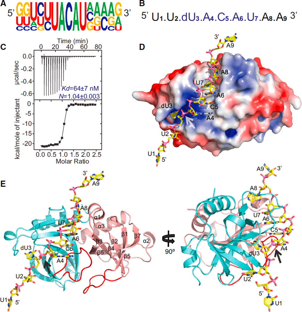
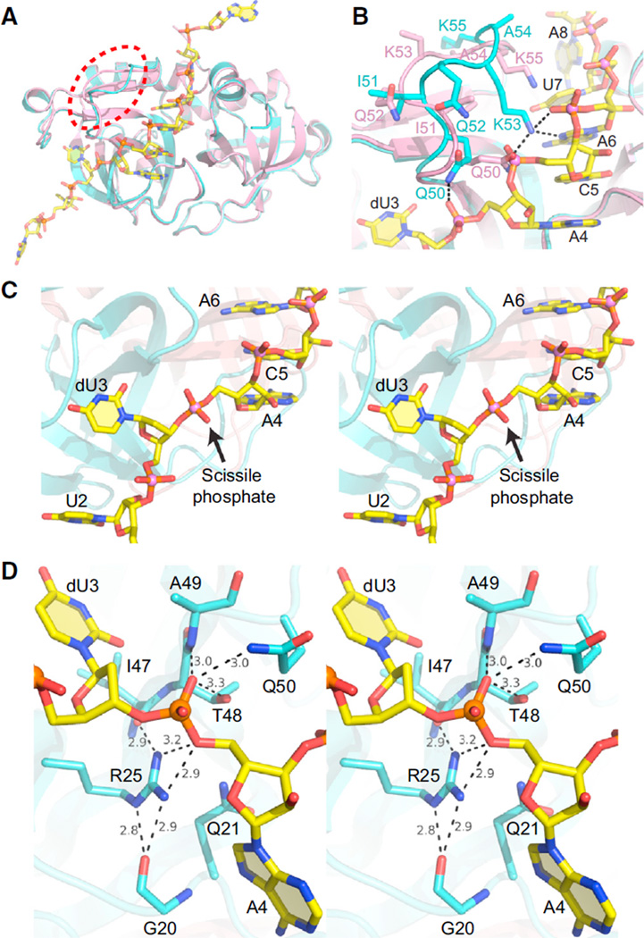
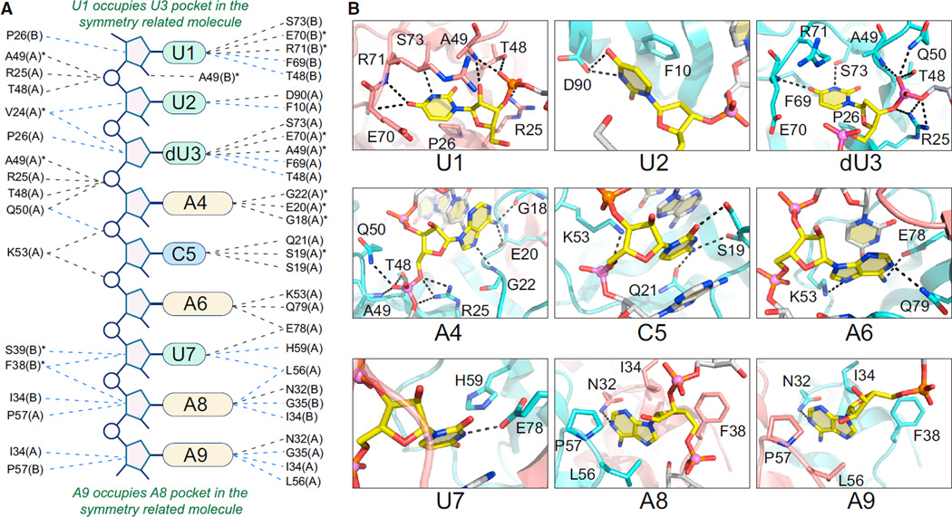
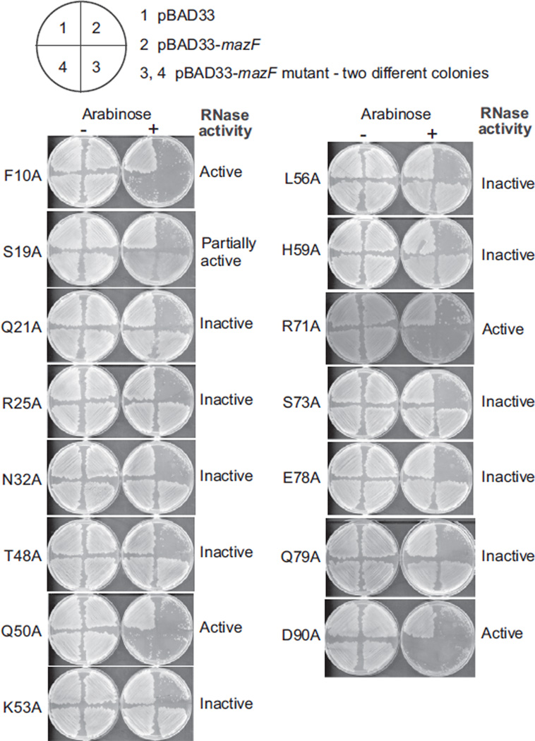
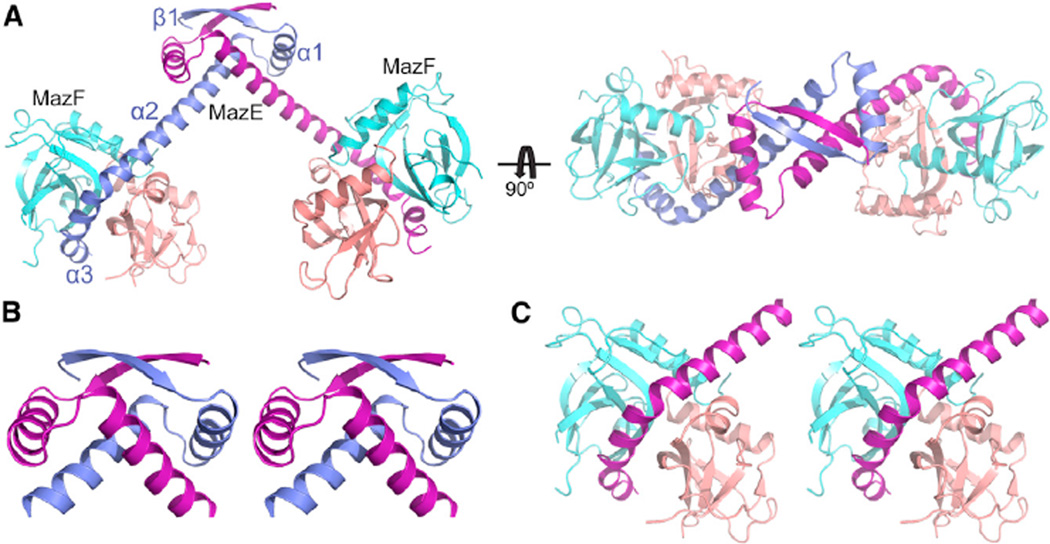
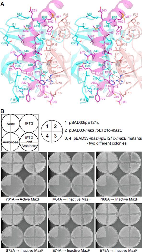
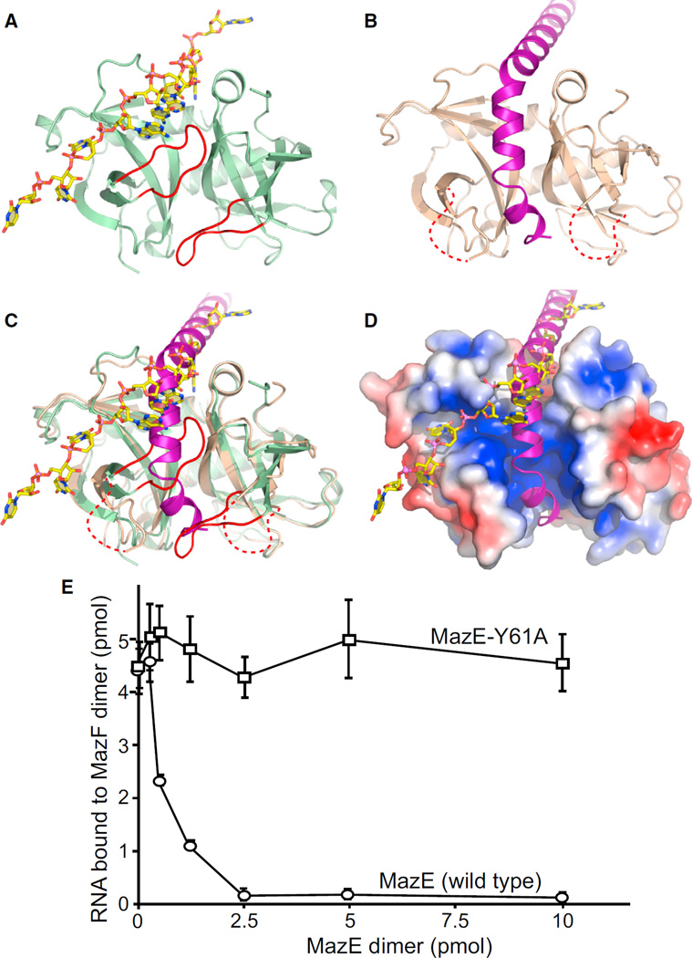
MazF is an mRNA interferase, which, upon activation during stress conditions, cleaves mRNAs in a sequence-specific manner, resulting in cellular growth arrest. During normal growth conditions, the MazF toxin is inactivated through binding to its cognate antitoxin, MazE. How MazF specifically recognizes its mRNA target and carries out cleavage and how the formation of the MazE-MazF complex inactivates MazF remain unclear. We present crystal structures of MazF in complex with mRNA substrate and antitoxin MazE in Bacillus subtilis. The structure of MazF in complex with uncleavable UUdUACAUAA RNA substrate defines the molecular basis underlying the sequence-specific recognition of UACAU and the role of residues involved in the cleavage through site-specific mutational studies. The structure of the heterohexameric (MazF)2-(MazE)2-(MazF)2 complex in Bacillus subtilis, supplemented by mutational data, demonstrates that the positioning of the C-terminal helical segment of MazE within the RNA-binding channel of the MazF dimer prevents mRNA binding and cleavage by MazF.


Figures

Figure 1
Figure 1. Binding Affinity and Overall Structure of B. subtilis MazF-RNA Complex Containing UACAU RNA Recognition Element
(A) Sequence logo plot of multiple sequence alignment of RNA sequences flanking the five different cleavage sites in MS2 RNA by MazF (Park et al., 2011). The height of the nucleotide corresponds to its conservation in the multiple-sequence alignment. (B) Sequence of the 9-mer RNA containing a dU at the first position of the U^ACAU RNA recognition element used for crystallization. (C) ITC measurement for binding of uncleavable 9-mer RNA (shown in B) containing UACAU RNA recognition element with MazF. The observed N value of 1.04 reflects 1 RNA bound per MazF dimer. (D) Electrostatic surface representation of the MazF protein in the MazF-RNA complex. Bound RNA is shown in stick representation and colored yellow. The arrow shows the position of scissile phosphate in the bound RNA. (E) Two views of the structure of the MazF-RNA complex. The MazF protein is shown in a ribbon representation, whereas the bound RNA (yellow) is shown in a stick representation. Two MazF subunits (cyan and salmon) form a symmetrical dimer. See also Figure S1 and Table S1.
Figure 2
Figure 2. Structural Comparison of B. subtilis MazF in Apo Form and in Complex with Bound RNA, Trajectory of the RNA in Bound Form, and the Active Site for Bound RNA Cleavage in B. subtilis MazF-RNA Complex
(A) Structural superposition of the apo form of MazF (pink) and MazF-RNA complex (cyan and yellow) showing conformational changes in the loop region between Gln50 and Lys55. The loop region is highlighted with the red ellipsoid. (B) Conformational changes in the loop formed by residues from Gln50 to Lys55 in the apo form of MazF (pink) and MazF-RNA complex (cyan and yellow). Side chain atoms are shown in stick representation. (C) Stereo view highlighting the changes in trajectory at the dU3-A4 step within the dUACAU segment of the bound RNA in the MazF-RNA complex. (D) Stereo view of the active site in the B. subtilis MazF-RNA complex showing that the residues present around the scissile phosphate present between dU3 and A4. The numbers list hydrogen bond distances between the bridging and nonbridging oxygens of the scissible phosphate and side chain and backbone atoms of amino acids of MazF lining the active site pocket.
Figure 3
Figure 3. Sequence-Specific Recognition of RNA Containing UACAU RNA Recognition Element by B. subtilis MazF
(A) Schematic representation of base-specific recognition of RNA substrate site by MazF. Dashed lines in black indicate hydrogen bonds, whereas dashed lines in blue show hydrophobic and van der Waal interactions between proteins and RNA. Asterisks designate interactions with main-chain atoms of the given amino acids, whereas A and B shown in parenthesis indicate two subunits of MazF. (B) Panels showing the recognition of individual nucleotides in RNA by MazF. Intermolecular hydrogen bonds are indicated by dashed black lines. For clarity, only one nucleotide is shown in yellow in each panel. See also Figures S2 and S3.
Figure 4
Figure 4. Impact of MazF Point Mutations for Residues Involved in RNA Binding and Cleavage in the B. subtilis MazF-RNA Complex
Expression of various MazF mutants under arabinose-inducible promoter (in duplicates in the bottom quadrants of the plate) was used to check the toxicity in E. coli cells. The upper left quadrant shows the expression of empty pBAD33 vector, whereas the upper right quadrant shows the expression of WT MazF as a control. Bacterial growth indicates a loss of toxicity, whereas the absence of bacterial growth shows behavior similar to WT MazF. See also Figure S4.
Figure 5
Figure 5. Structure of Heterohexameric B. subtilis MazE-MazF Complex and Overall Fold of B. subtilis MazE
(A) Ribbon representation of two views of the B. subtilis MazE-MazF complex. The two chains of MazF dimer are colored cyan and salmon, whereas the two chains of dimeric MazE are colored blue and purple. The hexameric complex is formed by two dimers of MazF bound to one dimer of MazE in the form of MazF2-MazE2-MazF2. (B) Stereo view of RHH motif present at the N-terminal end of MazE. (C) Stereo view of the region in MazE-MazF complex where C-terminal helices of MazE (purple) interact with MazF dimer. See also Figures S5 and S6 and Table S1.
Figure 6
Figure 6. Details of Intermolecular Protein-Protein Interaction Interface in B. subtilis MazE-MazF Complex and Impact of B. subtilis MazE Point Mutations on the Toxicity of B. subtilis MazE-MazF Complex in E. coli Cells
(A) Stereo view of the interface formed by residues from MazE and MazF in the B. subtilis complex. MazE is colored purple, whereas two chains in MazF dimer are colored cyan and salmon. Intermolecular hydrogen bonds are indicated by dashed black lines. (B) Effect of single amino acid mutations in B. subtilis MazE on the toxicity of B. subtilis MazE-MazF complex in E. coli cells. The expression of various MazE mutants (in duplicates in the bottom quadrants of the plate) under IPTG-inducible promoter along with WT MazF under arabinose-inducible promoter was used to check the toxicity in the E. coli cells. The upper left quadrants show the expression of empty pBAD33 and pET21c vector,whereas the upper right quadrants show the coexpression of WT MazF and MazE as a control. Bacterial growth indicates a loss of binding of MazE with MazF due to mutation in MazE, whereas the absence of bacterial growth shows behavior similar to WT MazF. See also Figure S7.
Figure 7
Figure 7. Structural Comparison of B. subtilis MazF-RNA and B. subtilis MazE-MazF Complexes Highlighting Overlap of Binding Site on the Dimeric Interface of MazF for Bound RNA and MazE and Competition Assay between MazE and ssRNA for MazF
(A) Structure of the MazF-RNA complex. A loop connecting β1 and β2 strands from each subunit and containing residues involved in RNA binding (highlighted in red) occupies part of the interface between the two subunits of MazF (light green). (B) Structure of the MazE-MazF complex. The C-terminal helices of MazE (purple) are positioned within the interface between the two subunits of MazF (light orange), thereby displacing the loop connecting β1 and β2 strands from each subunit, and these loops are disordered in the MazE-MazF complex. (C) Structural superposition of MazF-RNA complex and MazE-MazF complex showing overlap of binding sites for RNA and MazE on the dimeric interface of MazF. MazF is colored light green in the MazF-RNA complex, whereas it is colored light orange in the MazE-MazF complex. In the MazE-MazF complex, the C-terminal helical region of MazE (purple) displaces the loop connecting β1 and β2 strands from each subunit (highlighted in red), as seen in the MazF-RNA complex structure. (D) Electrostatic surface representation of MazF from the MazE-MazF complex with superposed RNA in the same view as in (C). (E) Filter-binding competition assay of MazF with ssRNA and MazE. A constant amount of MazF dimer (5 pmol) was incubated with variable amounts of WT MazE (circles) or MazE-Y61A (squares) on ice for 15 min. Then, labeled RNA (5 pmol) was added and incubated for 1 hr on ice. The amount of RNA bound to MazF was quantified with filter-binding assay. Error bars represent SEM (n = 3). See also Figure S7.

References

    1. Brown BL, Wood TK, Peti W, Page R. Structure of the Escherichia coli antitoxin MqsA (YgiT/b3021) bound to its gene promoter reveals extensive domain rearrangements and the specificity of transcriptional regulation. J. Biol. Chem. 2011;286:2285–2296. - PMC - PubMed
    1. Buts L, Lah J, Dao-Thi MH, Wyns L, Loris R. Toxin-antitoxin modules as bacterial metabolic stress managers. Trends Biochem. Sci. 2005;30:672–679. - PubMed
    1. Dalton KM, Crosson S. A conserved mode of protein recognition and binding in a ParD-ParE toxin-antitoxin complex. Biochemistry. 2010;49:2205–2215. - PMC - PubMed
    1. De Jonge N, Garcia-Pino A, Buts L, Haesaerts S, Charlier D, Zangger K, Wyns L, De Greve H, Loris R. Rejuvenation of CcdB-poisoned gyrase by an intrinsically disordered protein domain. Mol. Cell. 2009;35:154–163. - PubMed
    1. Gerdes K, Christensen SK, Løbner-Olesen A. Prokaryotic toxin-antitoxin stress response loci. Nat. Rev. Microbiol. 2005;3:371–382. - PubMed

Publication types

MeSH terms

LinkOut - more resources