. 2013 Nov 21;52(4):574-82.
doi: 10.1016/j.molcel.2013.09.018. Epub 2013 Oct 10.

The translational landscape of the mammalian cell cycle

Affiliations

The translational landscape of the mammalian cell cycle

Craig R Stumpf et al. Mol Cell. .

Abstract

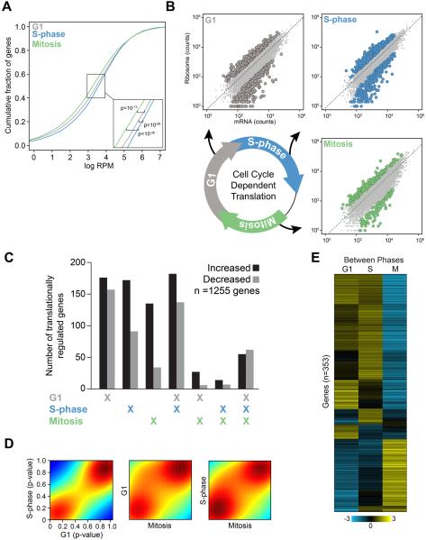
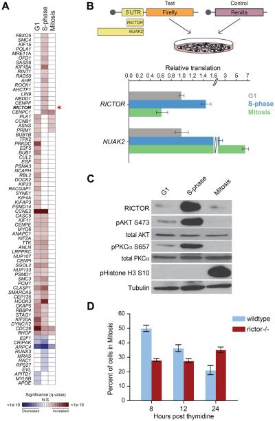
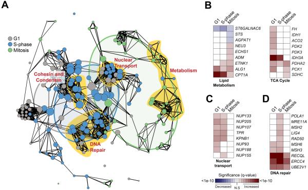
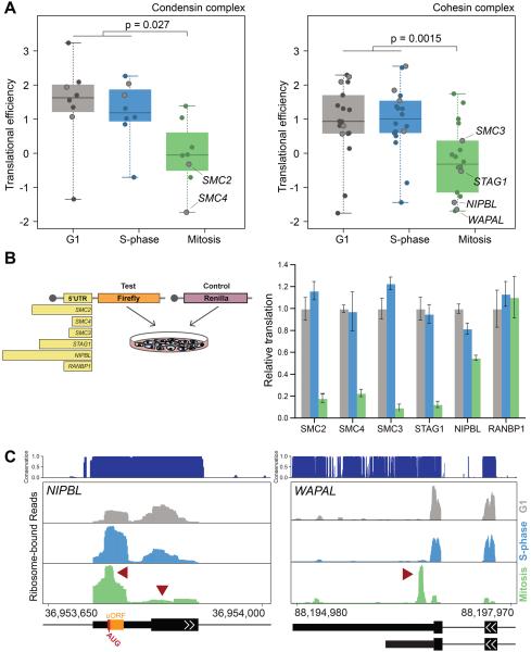
Gene regulation during cell-cycle progression is an intricately choreographed process, ensuring accurate DNA replication and division. However, the translational landscape of gene expression underlying cell-cycle progression remains largely unknown. Employing genome-wide ribosome profiling, we uncover widespread translational regulation of hundreds of mRNAs serving as an unexpected mechanism for gene regulation underlying cell-cycle progression. A striking example is the S phase translational regulation of RICTOR, which is associated with cell cycle-dependent activation of mammalian target of rapamycin complex 2 (mTORC2) signaling and accurate cell-cycle progression. We further identified unappreciated coordination in translational control of mRNAs within molecular complexes dedicated to cell-cycle progression, lipid metabolism, and genome integrity. This includes the majority of mRNAs comprising the cohesin and condensin complexes responsible for maintaining genome organization, which are coordinately translated during specific cell cycle phases via their 5' UTRs. Our findings illuminate the prevalence and dynamic nature of translational regulation underlying the mammalian cell cycle.


Figures

Figure 1
Figure 1. Systematic and multifaceted translational control of gene expression during the mammalian cell cycle.
(A) The cumulative fraction of ribosome-bound mRNA on all expressed transcripts in each phase of the cell cycle is shown as a function of increasing ribosome-bound mRNA. X-axis represents the scaled fraction of total ribosome-bound reads and the Y-axis represents the fraction of expressed transcripts. (B) Representative scatter plots illustrate ribosome occupancy as a function of mRNA abundance (measured as sequencing read counts). The dashed line represents the expected level of ribosome occupancy given mRNA abundance (see Supplemental Experimental Procedures). mRNAs with statistically significant translational regulation are those with greater or less than the expected levels of ribosome occupancy given their mRNA expression (FDR < 1%; G1 is grey, S-phase is blue, and Mitosis is green). (C) The total number of genes with significantly increased (black) or decreased (grey) ribosome occupancy are shown, including those unique to a given phase or shared between multiple phases of the cell cycle. (D) Density plots of the nominal p-value of ribosome occupancy for each gene between any two phases of the cell cycle (colors designate the density of genes in a given region where red and blue are the most and least dense respectively). (E) Unsupervised hierarchical clustering of mRNA translation across the phases of the cell cycle using the 353 differentially translationally regulated transcripts between any two phases in direct comparisons. Genes and cell cycle phases are clustered based on the level of normalized ribosome occupancy (mean-centered translational efficiency by gene, scales and colors indicate the direction and magnitude of mRNA translation S is S phase, M is Mitosis). See also Figure S1, Tables S1-2.
Figure 2
Figure 2. Phase-dependent translational control of key cell cycle regulators including RICTOR and mTOR signaling.
(A) A heat map of the significance of translational regulation among cell cycle progression genes (asterisk: RICTOR). Shading represents significance level of increased or decreased translational regulation (red and blue, as indicated). (B) A diagram describing the 5′ UTR luciferase reporter assay (top panel). 5′ UTRs cloned into the firefly reporter are scaled to length. Capped mRNAs are transfected into synchronized cells prior to assaying reporter levels. Levels of translation directed by RICTOR or NUAK2 5′UTRs are shown: Y-axis is reporter value relative to cells in G1 (bottom panel). (C) Western blots showing RICTOR protein levels and phosphorylation of mTORC2 targets (AKT-S473, PKCα-S657). pHistone H3 is a mitosis marker. Tubulin is a loading control. (D) Mitotic progression of thymidine-synchronized MEFs: Y-axis is percent mitotic cells. Bars represent the mean +/- SD. See also Figure S2.
Figure 3
Figure 3. Translational co-regulation of large molecular complexes during cell cycle progression.
(A) Among translationally regulated genes during the cell cycle, a network of statistically significant functional enrichments (nodes, nominal p-value < 0.05; radius scaled to the number of genes) and their relatedness (edges, spearman correlation ρ ≥ 0.3) indicate a highly interconnected set of modules of molecular function. Groups of nodes closely related by function are highlighted in yellow and labeled. A Venn diagram overlay represents the relative overlap of enriched molecular function between cell cycle phases (G1 is grey, S-phase is blue, and Mitosis is green). Heatmaps highlight the significance of translational regulation of genes that define representative functional categories including lipid metabolism and TCA cycle (B), nuclear transport (C), and DNA repair (D). Shading represents significance level of increased or decreased translational regulation (red and blue, as indicated). See also Table S3.
Figure 4
Figure 4. Condensin and cohesion complex components are coordinately translationally regulated during the cell cycle at the level of their 5′UTR.
(A) The translational efficiency of components of the condensin (left) and cohesin (right) complexes during each cell cycle phase are indicated (specific genes mentioned elsewhere are outlined for clarity). (B) A diagram of the luciferase reporter assay with 5′UTRs scaled to length (left). Levels of translation directed by specific 5′ UTRs are indicated: Y-axis is reporter value relative to cells in G1 (right). Bars represent mean +/- SD from 6 replicates. (C) The level of bound ribosome in the 5′ UTR of NIPBL (left) and the 3′ UTR of WAPAL (right), with peaks of interest denoted by red arrows (evolutionary conservation is indicated; G1 is grey, S-phase is blue, and Mitosis is green). Representative gene features are indicated: uORF is designated by an orange box, narrow or wide black bars represent UTRs and coding exons respectively, black lines indicate introns, and arrows indicate the coding strand. Numbers represent absolute genomic positions. See also Figure S3.

References

    1. Alessi DR, Andjelkovic M, Caudwell B, Cron P, Morrice N, Cohen P, Hemmings BA. Mechanism of activation of protein kinase B by insulin and IGF-1. EMBO J. 1996;15:6541–6551. - PMC - PubMed
    1. Alessi DR, James SR, Downes CP, Holmes AB, Gaffney PR, Reese CB, Cohen P. Characterization of a 3-phosphoinositide-dependent protein kinase which phosphorylates and activates protein kinase Balpha. Curr Biol. 1997;7:261–269. - PubMed
    1. Antonin W, Ellenberg J, Dultz E. Nuclear pore complex assembly through the cell cycle: regulation and membrane organization. FEBS Lett. 2008;582:2004–2016. - PubMed
    1. Borck G, Zarhrate M, Cluzeau C, Bal E, Bonnefont JP, Munnich A, CormierDaire V, Colleaux L. Father-to-daughter transmission of Cornelia de Lange syndrome caused by a mutation in the 5′ untranslated region of the NIPBL Gene. Hum Mutat. 2006;27:731–735. - PubMed
    1. Bose T, Lee KK, Lu S, Xu B, Harris B, Slaughter B, Unruh J, Garrett A, McDowell W, Box A, et al. Cohesin proteins promote ribosomal RNA production and protein translation in yeast and human cells. PLoS Genet. 2012;8:e1002749. - PMC - PubMed

Publication types

MeSH terms