. 2013 Nov 5;18(5):649-59.
doi: 10.1016/j.cmet.2013.09.008. Epub 2013 Oct 10.

Exercise induces hippocampal BDNF through a PGC-1α/FNDC5 pathway

Affiliations

Exercise induces hippocampal BDNF through a PGC-1α/FNDC5 pathway

Christiane D Wrann et al. Cell Metab. .

Abstract

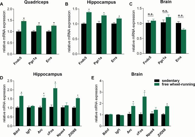
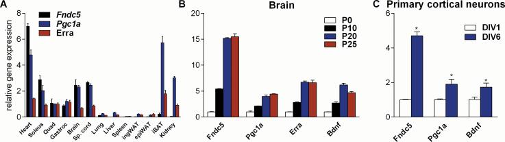
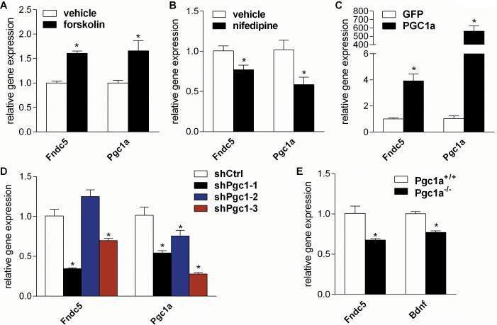
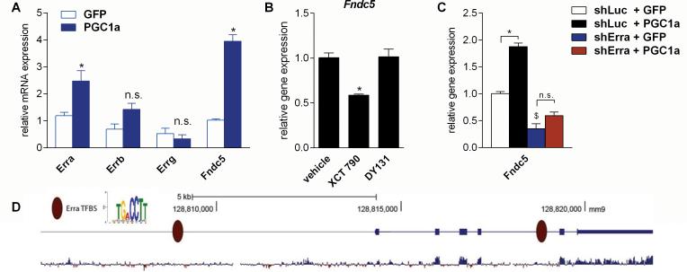
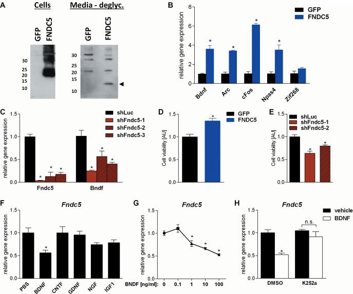
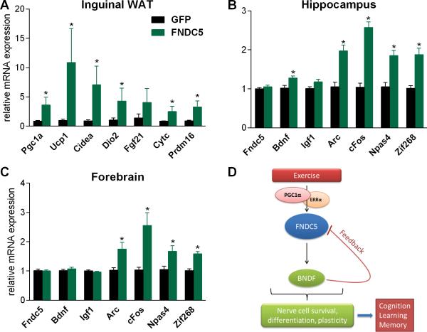
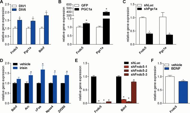
Exercise can improve cognitive function and has been linked to the increased expression of brain-derived neurotrophic factor (BDNF). However, the underlying molecular mechanisms driving the elevation of this neurotrophin remain unknown. Here we show that FNDC5, a previously identified muscle protein that is induced in exercise and is cleaved and secreted as irisin, is also elevated by endurance exercise in the hippocampus of mice. Neuronal Fndc5 gene expression is regulated by PGC-1α, and Pgc1a(-/-) mice show reduced Fndc5 expression in the brain. Forced expression of FNDC5 in primary cortical neurons increases Bdnf expression, whereas RNAi-mediated knockdown of FNDC5 reduces Bdnf. Importantly, peripheral delivery of FNDC5 to the liver via adenoviral vectors, resulting in elevated blood irisin, induces expression of Bdnf and other neuroprotective genes in the hippocampus. Taken together, our findings link endurance exercise and the important metabolic mediators, PGC-1α and FNDC5, with BDNF expression in the brain.


Figures

Figure 1
Figure 1. Endurance exercise induces hippocampal Fndc5 gene expression
(A-E) Male six week old C57/Bl6 wild type mice were individually housed in cages with access to a running-wheel (free wheel-running) or without (sedentary). Mice were exercised for 30 days and sacrificed approximately 10 h after their last bout of exercise. Data are shown as mRNA levels relative to Rsp18 expression, expressed as mean ± SEM. *P < 0.05 compared to sedentary control group. See also Figure S1.
Figure 2
Figure 2. Fndc5 gene expression correlates with Pgc1a expression levels in various tissues and developmental stages
(A) The indicated tissues were harvested from male 13 week old C57/Bl6 wild type mice. Quad = quadriceps muscle, Gastroc = gastrocnemius muscle, Sp. Cord = spinal cord, ingWAT = inguinal white adipose tissue, epiWAT = epididymal white adipose tissue, iBAT = interscapular brown adipose tissue. (B) Brains were harvested from C57/Bl6 wild type mice at the indicated postnatal (P) time points. (C) Primary cortical neurons were isolated from C57/Bl6 wild type E17 embryos and cultured for the indicated days in vitro. mRNA was prepared and gene expression was assessed by qPCR. All data are shown as mRNA levels relative to Rsp18 expression, expressed as mean ± SEM. *P < 0.05 compared to control group.
Figure 3
Figure 3. Neuronal Fndc5 gene expression is regulated by a PGC-1α
(A) Primary cortical neurons at DIV 7 were treated with either forskolin (10 μM), a stimulator intracellular cAMP levels, or vehicle for overnight. (B) Primary cortical neurons at DIV 7 were treated with nifedipine (5 μM), a L-type calcium channel blocker, or vehicle for overnight. (C) Primary cortical neurons at DIV 7 were transduced with either PGC-1α or GFP adenovirus and harvested 48 hrs later. (D) Primary cortical neurons at DIV 5 were transduced with lentivirus carrying the specified shRNA hairpins against Pgc1a or luciferase (Luc) as control and were harvested four days later. (E) Cortices were harvested from either male five months old Pgc1a KO (Pgc1a−/−) or wild type mice (Pgc1a+/+). mRNA was prepared and gene expression was assessed by qPCR. All data are shown as mRNA levels relative to Rsp18 expression, expressed as mean ± SEM. *P < 0.05 compared to corresponding control group. See also Figure S2.
Figure 4
Figure 4. ERRα is a key interacting transcription factor with PGC-1α for regulating Fndc5 gene expression in neurons
(A) Primary cortical neurons at DIV 7 were transduced with either PGC-1α or GFP adenovirus and harvested 48 hrs later. *P < 0.05 compared to control group. (B) Primary cortical neurons at DIV 7 were treated with either XCT 790 (1 μM), a selective inverse ERRα agonist, DY131 (1 μM), a selective ERRβ and ERRγ agonist, or vehicle for overnight. *P < 0.05 compared to vehicle only group. (C) Primary cortical neurons at DIV 4 were transduced with lentivirus carrying shRNA hairpins against either Erra or luciferase (Luc) as control. Three days later were cells were transduced with either PGC-1α or GFP adenovirus and harvested 48 hrs later. mRNA was prepared and gene expression was assessed by qPCR. Data are shown as mRNA levels relative to Rsp18 expression, expressed as mean ± SEM. *P < 0.05 compared to corresponding shLuc expressing control group. $P < 0.05 compared to corresponding GFP expressing control group. Data (A-C) are shown as mRNA levels relative to Rsp18 expression, expressed as mean ± SEM. (D) Analysis of the murine Fndc5 promoter for putative ERREs. The murine Fndc5 gene and 6 kb of its upstream promoter were searched for the canonical ERRE: TGA CCTT. Genomic coordinates are given according to the assembly mm9 from the UCSC Genome Browser. The bottom diagram indicates the degree of mammalian conservation across the genomic locus. The presented motif was modified from www.factorbook.org (Wang et al., 2012). See also Figure S3.
Figure 5
Figure 5. FNDC5 regulates Bdnf gene expression in a cell-autonomous manner and recombinant BDNF decreases Fndc5 gene expression as part of negative feedback loop
(A) Primary cortical neurons at DIV 6 were transduced with either FNDC5 or GFP adenovirus. Whole cell lysates and conditioned media were harvested and analyzed by immunoblotting. Intensity of unspecific bands and Ponceau staining were used to assess equal loading. deglyc. = deglycosylation. (B) Primary cortical neurons at DIV 7 were transduced with either FNDC5 or GFP adenovirus. Forty-eight hours later mRNA was prepared and gene expression was assessed by qPCR. (C) Primary cortical neurons at DIV 5 were transduced with lentivirus carrying the specified shRNA hairpins against Fndc5 or luciferase (Luc) as control. Four days later mRNA was prepared and gene expression was assessed by qPCR. (D) Primary cortical neurons at DIV 7 were transduced with either FNDC5 or GFP adenovirus. Cell viability was assessed three days later using the CellTiter-Glo® Luminescent Cell Viability Assay. AU = arbitrary unit. (E) Primary cortical neurons at DIV 5 were transduced with lentivirus carrying the specified shRNA hairpins against Fndc5 or luciferase (Luc) as control. Cell viability was assessed three days later using the CellTiter-Glo® Luminescent Cell Viability Assay. AU = arbitrary unit. (F) Primary cortical neurons at DIV 7 were stimulated with the indicated recombinant neurotrophins and growth factors (100ng/ml) for overnight. mRNA was prepared and gene expression was assessed by qPCR. (G) Primary cortical neurons at DIV 7 were stimulated with human recombinant BDNF at the indicated concentrations or vehicle for overnight. mRNA was prepared and gene expression was assessed by qPCR. (H) Primary cortical neurons at DIV 6 were treated either with the TrkB inhibitor K252a (50nM) or vehicle. Twenty-four hours later human recombinant BDNF (100ng/ml) or vehicle was added for overnight stimulation. mRNA was prepared and gene expression was assessed by qPCR. Data (B, C and F- H) are shown as mRNA levels relative to Rsp18 expression. All data are expressed as mean ± SEM. *P < 0.05 compared to corresponding control group.
Figure 6
Figure 6. Peripheral delivery of FNDC5 by adenoviral vectors increases Bdnf expression in the hippocampus
(A-C) Five week old male wild-type BALB/c mice were injected with GFP- or FNDC5-expressing adenoviral particles intravenously. Animals were sacrificed seven days later and (A) inguinal/subcutaneous fat pads (WAT=white adipose tissues), (B) hippocampus, and forebrain (C) were collected and mRNA was prepared and gene expression was assessed by qPCR. Data are shown as mRNA levels relative to Rsp18 expression, expressed as mean ± SEM. *P < 0.05 compared to wild type control group. (D) Model of the hippocampal PGC-1α/FNDC5/BDNF pathway in exercise. Endurance exercise stimulates increases hippocampal Fndc5 gene expression through a PGC-1α/Errα transcriptional complex. This elevated Fndc5 gene expression stimulates in turn Bdnf gene expression. BDNF is the master regulator of nerve cell survival, differentiation and plasticity in the brain. This will lead to improved cognitive function, learning and memory, which are known beneficial effects of exercise on the brain. See also Figure S4.
Figure 7
Figure 7. PGC-1α/FNDC5/BDNF pathway in primary hippocampal neurons
(A) Primary hippocampal neurons were isolated from C57/Bl6 wild type E17 embryos and cultured for the indicated days in vitro. (B) Primary hippocampal neurons at DIV 7 were transduced with either PGC-1α or GFP adenovirus and harvested 48 hrs later. (C) Primary hippocampal neurons at DIV 5 were transduced with lentivirus carrying the specified shRNA hairpin against Pgc1a or luciferase (Luc) as control and harvested four days later. (D) Primary hippocampal neurons were stimulated with recombinant irisin (1ug/ml) at DIV 5 and 6 and harvested 24 hrs later (E) Primary hippocampal neurons at DIV 5 were transduced with lentivirus carrying the specified shRNA hairpins against Fndc5 or luciferase (Luc) as control and harvested four days later. (F) Primary hippocampal neurons at DIV 7 were stimulated with recombinant BDNF (100ng/ml) for overnight. mRNA was prepared and gene expression was assessed by qPCR. Data are shown as mRNA levels relative to Rsp18 expression, expressed as mean ± SEM. *P < 0.05 compared to corresponding control group. See also Figure S5.

Comment in

  • BDNF (I)rising from exercise.
    Xu B. Xu B. Cell Metab. 2013 Nov 5;18(5):612-4. doi: 10.1016/j.cmet.2013.10.008. Cell Metab. 2013. PMID: 24206660 Free PMC article.

References

    1. Ahlskog JE. Does vigorous exercise have a neuroprotective effect in Parkinson disease? Neurology. 2011;77:288–294. - PMC - PubMed
    1. Arany Z, Foo SY, Ma Y, Ruas JL, Bommi-Reddy A, Girnun G, Cooper M, Laznik D, Chinsomboon J, Rangwala SM, et al. HIF-independent regulation of VEGF and angiogenesis by the transcriptional coactivator PGC-1alpha. Nature. 2008;451:1008–1012. - PubMed
    1. Arida RM, Cavalheiro EA, da Silva AC, Scorza FA. Physical activity and epilepsy: proven and predicted benefits. Sports medicine (Auckland, NZ) 2008;38:607–615. - PubMed
    1. Bartlett WP, Banker GA. An electron microscopic study of the development of axons and dendrites by hippocampal neurons in culture. I. Cells which develop without intercellular contacts. J Neurosci. 1984;4:1944–1953. - PMC - PubMed
    1. Bostrom P, Wu J, Jedrychowski MP, Korde A, Ye L, Lo JC, Rasbach KA, Bostrom EA, Choi JH, Long JZ, et al. A PGC1-alpha-dependent myokine that drives brown-fat-like development of white fat and thermogenesis. Nature. 2012 - PMC - PubMed

Publication types

MeSH terms

Substances