. 2014 Jan;80(1):77-85.
doi: 10.1128/AEM.02310-13. Epub 2013 Oct 11.

Characterization of bacteriophages Cp1 and Cp2, the strain-typing agents for Xanthomonas axonopodis pv. citri

Affiliations

Characterization of bacteriophages Cp1 and Cp2, the strain-typing agents for Xanthomonas axonopodis pv. citri

Abdelmonim Ali Ahmad et al. Appl Environ Microbiol. 2014 Jan.

Abstract

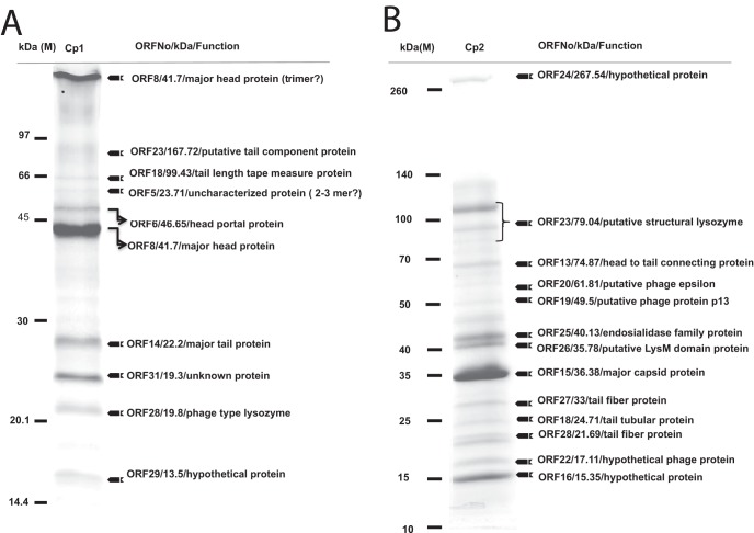
The strains of Xanthomonas axonopodis pv. citri, the causative agent of citrus canker, are historically classified based on bacteriophage (phage) sensitivity. Nearly all X. axonopodis pv. citri strains isolated from different regions in Japan are lysed by either phage Cp1 or Cp2; Cp1-sensitive (Cp1(s)) strains have been observed to be resistant to Cp2 (Cp2(r)) and vice versa. In this study, genomic and molecular characterization was performed for the typing agents Cp1 and Cp2. Morphologically, Cp1 belongs to the Siphoviridae. Genomic analysis revealed that its genome comprises 43,870-bp double-stranded DNA (dsDNA), with 10-bp 3'-extruding cohesive ends, and contains 48 open reading frames. The genomic organization was similar to that of Xanthomonas phage phiL7, but it lacked a group I intron in the DNA polymerase gene. Cp2 resembles morphologically Escherichia coli T7-like phages of Podoviridae. The 42,963-bp linear dsDNA genome of Cp2 contained terminal repeats. The Cp2 genomic sequence has 40 open reading frames, many of which did not show detectable homologs in the current databases. By proteomic analysis, a gene cluster encoding structural proteins corresponding to the class III module of T7-like phages was identified on the Cp2 genome. Therefore, Cp1 and Cp2 were found to belong to completely different virus groups. In addition, we found that Cp1 and Cp2 use different molecules on the host cell surface as phage receptors and that host selection of X. axonopodis pv. citri strains by Cp1 and Cp2 is not determined at the initial stage by binding to receptors.


Figures

FIG 1
FIG 1
Single-step growth curves for Cp1 (A) and Cp2 (B). The PFU per infected cell in cultures of MAFF 301080 for Cp1 and MAFF 673010 for Cp2 at different times postinfection are shown. Samples were taken at intervals (every 10 min up to 3.5 h for Cp1 and every 30 min up to 5 h for Cp2). Error bars indicate standard deviations (n = 3).
FIG 2
FIG 2
Genomic organization of phages Cp1 (A) and phiL7 (B). Colored arrows indicate the directions and categories of the genes. Broken arrows and black knobs indicate the σ70-type promoters and predicted terminators for transcription, respectively. “No similarity” was determined using an E value lower than e−4 as a cutoff for notable similarity.
FIG 3
FIG 3
Comparison of amino acid sequences of RNA polymerases (RNAP) encoded by Xanthomonas phages. (A) The amino acid sequence of Cp1 ORF33 (AB720063) was aligned with those of phiL7 p35 (ACE75775.1)., Xop411 p32 (ABK00180.1), Xp10 32L (AAP58699.1), OP1 ORF33 (BAE72738.1), and coliphage T7 RNAP (NP_041960.1) using ClustalX. The ClustalX coloring scheme depends on both the residue type and the pattern of conservation within a column (http://www.cgl.ucsf.edu/chimera/docs/ContributedSoftware/multalignviewer/cxcolor.html). Conservation scores are drawn below the alignment. (B) The unrooted dendrogram was constructed with the Treeview tool using the maximum likelihood method based on a complete protein sequence alignment of RNAP proteins from other phages.
FIG 4
FIG 4
Genomic organization of phages of Cp2 (A) and Escherichia coli T7 (B). Arrows indicate the sizes and directions of the genes. The typical podovirus genome represented by T7 phage consists of three functional modules: class I, class II, and class III, as shown in panel B (35). Putative class I (white), class II (white), and class III (red) modules for Cp2 are shown in panel A.
FIG 5
FIG 5
Proteomic analysis of Cp1 (A) and Cp2 (B) particles. Proteins of purified phage particles were separated in 10% (wt/vol) polyacrylamide gel by SDS-PAGE and stained with Coomassie brilliant blue. The protein bands were recovered, digested in gel, and subjected to LC-MS/MS analysis. On the right are the descriptions of the genes, their deduced molecular sizes based on the ORF sequences, and their possible functions. Positions of size markers are shown on the left. For some protein bands, possible oligomerization of the phage protein was observed.
FIG 6
FIG 6
Attachment of SYBR gold-labeled phage particles to bacterial cells and staining of the cells by possible injection of phage DNA. (A) Cp1 particles were added to cells of host strain MAFF 301080; (B) Cp2 particles were added to cells of host strain MAFF 673010. Cells from panels A and B were observed under a fluorescence microscope at 10 min (MOI = 1, left panel) and 20 min (MOI = 10, right panel) postinfection. The attachment of the phage particles to the bacterial cell surface is shown. Phage particles appear as tiny spots (arrows). At 10 min postinfection, bacteria cell were not stained (bright and dark fields), while cells became stained at 20 min postinfection (dark field). Cp1 (C) and Cp2 (D) particles were also added to cells of MAFF 301080 (Cp1s/Cp2r), MAFF 673010 (Cp1r/Cp2s), and MAFF 311130 (Cp1r/Cp2r). Cells were observed under a fluorescence microscope at 30 min postinfection. Bacterial cells were stained by SYBR gold, indicating injection of phage DNA into the Cp1s/Cp2r or Cp1r/Cp2s as well as Cp1r/Cp2r cells. Bar, 10 μm.

References

    1. Civerelo EL. 1984. Bacterial canker disease of citrus. J. Rio. Grande Val. Hort. Soc. 37:127–145
    1. Gottwald TR, Graham JH, Schubert TS. 2002. Citrus canker: the pathogen and its impact. Plant health progress. http://www.plantmanagementnetwork.org/pub/php/review/citruscanker/
    1. Stall RE, Civerolo EL. 1991. Research relating to the recent outbreak of citrus canker in Florida. Annu. Rev. Phytopathol. 29:399–420 - PubMed
    1. Wakimoto S. 1967. Some characteristics of citrus canker bacteria, Xanthomonas citri (Hasse) Dowson, and the related phages isolated from Japan. Ann. Phytopathol. Soc. Jpn. 33:301–310
    1. Obata T. 1974. Distribution of Xanthomonas citri strain in relation to the sensitivity to phage CP1 and CP2. Ann. Phytopathol. Soc. Jpn. 40:6–13

Associated data