. 2014 Apr;39(4):1017.
doi: 10.1002/jmri.24224. Epub 2013 Oct 7.

Registration of dynamic contrast-enhanced MRI of the common carotid artery using a fixed-frame template-based squared-difference method

Affiliations

Registration of dynamic contrast-enhanced MRI of the common carotid artery using a fixed-frame template-based squared-difference method

Sarayu Ramachandran et al. J Magn Reson Imaging. 2014 Apr.

Abstract

Purpose: This study examines template-based squared-difference registration for motion correction in dynamic contrast-enhanced (DCE) MRI studies of the carotid artery wall and compares the results of fixed-frame template-based registration with a previously proposed consecutive-frame registration method.

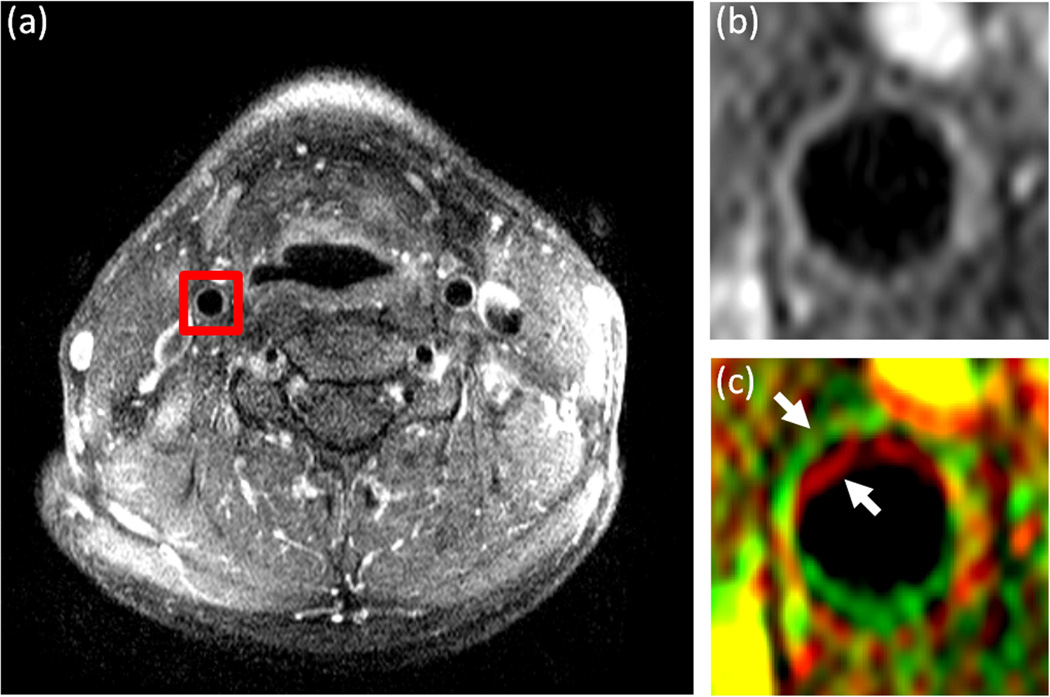
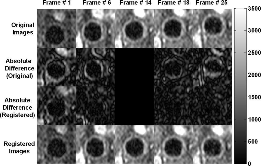
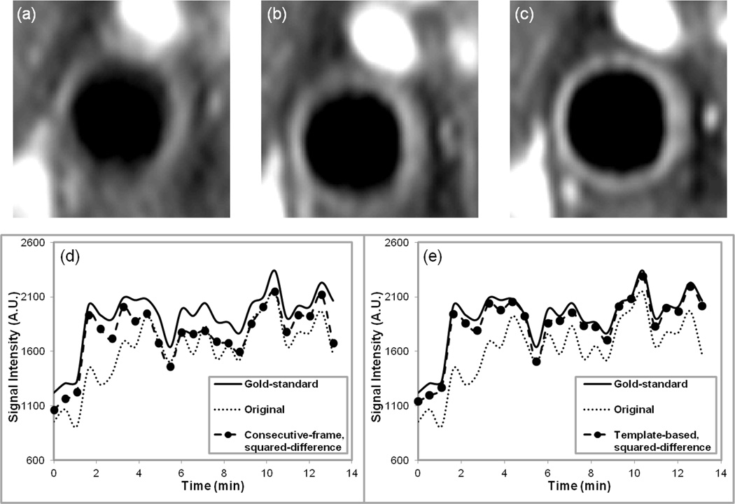
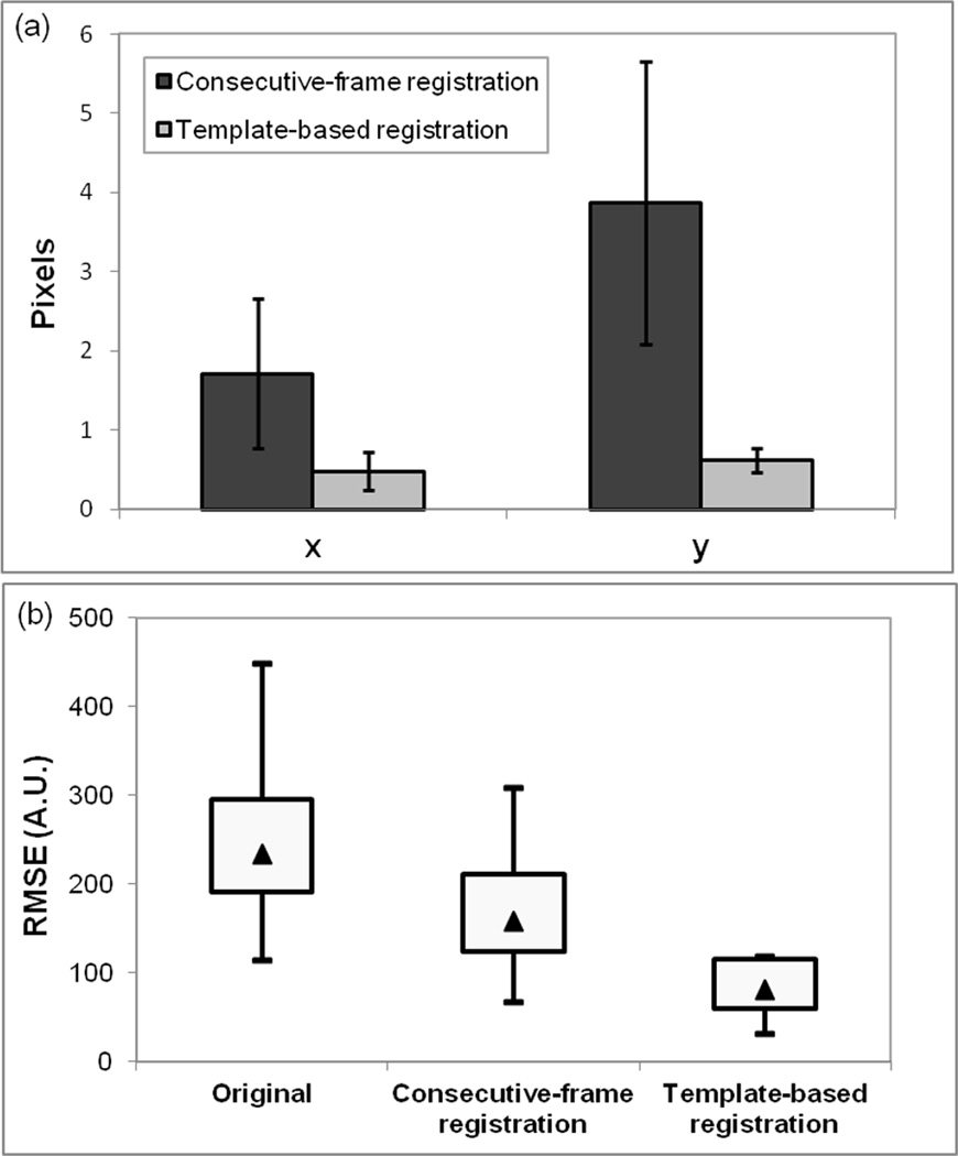
Materials and methods: Ten T1-weighted black-blood, turbo spin-echo DCE-MRI studies of the carotid artery wall were used to test template-based squared-difference registration. An intermediate image from each series was selected as the fixed-frame template for registration. Squared-difference minimization was used to align each image and template. Time-intensity curves generated from data aligned with template-based squared-difference registration were compared with gold standard curves created by drawing regions of interest on each image in the series. The results were also compared with unregistered data and data after consecutive-frame squared-difference registration.

Results: An analysis of variance test of root mean-square error values between gold standard curve and curves from unregistered data and data registered with consecutive-frame and fixed-frame template-based methods was significant (P < 0.005) with template-based squared-difference registration producing curves that most closely matched the gold standard.

Conclusion: A fixed-frame template-based squared-difference registration method was proposed and validated for alignment of DCE-MRI of carotid arteries.

Keywords: registration; template-matching; vascular DCE-MRI.


Figures

Figure 1
Figure 1
DCE-MR images of the carotid arteries in the neck of a representative subject. a: A single image from a DCE-MRI series with a red square surrounding the right carotid artery indicating the cropped region used for analysis. b: The 14th frame in the DCE-MRI series used as the template image for registration and (c) the 15th frame in the series (rendered in green) overlaid on the 14th frame (rendered in red). The two consecutive frames are overlaid to show the misalignment between images in the series before registration. The arrows indicate the position of the carotid artery wall in each of the frames.
Figure 2
Figure 2
Several frames from a DCE-MRI series of a typical subject are presented to illustrate the motion of the carotid artery over the course of the sequence acquisition and the need for registration. In the original images (top row), the vessel changes position from near the base of the image in frame # 1 to closer to the top in frame # 25. In the registered images (bottom row), the position of the vessel remains relatively stable in all the frames. Frame # 14 was the template image for this subject (middle column). The second row shows the absolute difference between the original images and the template image, and the third row shows the corresponding difference between the registered images and the template. After registration, there is much less difference between the various frames and the template, indicating that the images were aligned appropriately. All images are displayed with the same intensity levels for comparison.
Figure 3
Figure 3
Average images and time-intensity curves. Images obtained after averaging together all 25 frames in a DCE-MRI series (a) original data, before registration, (b) after registration with the consecutive-frame squared-difference method, (c) after registration with the template-based squared-difference method. All images are displayed with the same intensity levels for comparison. Both plots in (d) and (e) display the gold-standard and original data time-intensity curves along with the consecutive- frame curve in (d) and the template-based time-intensity curve in (e).
Figure 4
Figure 4
a: Column graph showing the average absolute registration errors over the subject population in the x- and y-directions for consecutive-frame and template-based registration. b: Box plot of the average RMSE values over the test population before registration, and after registration with consecutive-frame and template-based methods. The bottom and top of each box represents the 25th and 75th percentile of RMSE values respectively and the ends of the whiskers in the box plot represent the maximum and minimum RMSE values for each method. The triangle in each box represents the median RMSE value for the method.

References

    1. Moreno PR, Purushothaman KR, Fuster V, et al. Plaque neovascularization is increased in ruptured atherosclerotic lesions of human aorta: implications for plaque vulnerability. Circulation. 2004;110:2032–2038. - PubMed
    1. Jain RK, Finn AV, Kolodgie FD, Gold HK, Virmani R. Antiangiogenic therapy for normalization of atherosclerotic plaque vasculature: a potential strategy for plaque stabilization. Nat Clin Pract Cardiovasc Med. 2007;4:491–502. - PubMed
    1. Langheinrich AC, Kampschulte M, Buch T, Bohle RM. Vasa vasorum and atherosclerosis - Quid novi? Thromb Haemost. 2007;97:873–879. - PubMed
    1. Kerwin WS, O’Brien KD, Ferguson MS, Polissar N, Hatsukami TS, Yuan C. Inflammation in carotid atherosclerotic plaque: a dynamic contrast-enhanced MR imaging study. Radiology. 2006;241:459–468. - PMC - PubMed
    1. Gaens ME, Backes WH, Rozel S, et al. Dynamic contrast-enhanced MR imaging of carotid atherosclerotic plaque: model selection, reproducibility, and validation. Radiology. 2013;266:271–279. - PubMed

Publication types

LinkOut - more resources