Review
. 2014 Apr 10;20(11):1754-69.
doi: 10.1089/ars.2013.5666. Epub 2013 Dec 10.

Coupling heme and iron metabolism via ferritin H chain

Affiliations
Review

Coupling heme and iron metabolism via ferritin H chain

Raffaella Gozzelino et al. Antioxid Redox Signal. .

Abstract

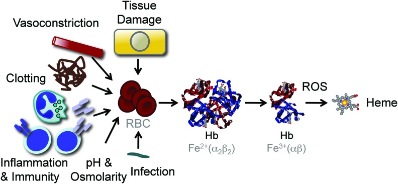
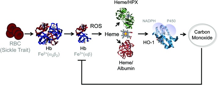
Significance: Inflammation and immunity can be associated with varying degrees of heme release from hemoproteins, eventually leading to cellular and tissue iron (Fe) overload, oxidative stress, and tissue damage. Presumably, these deleterious effects contribute to the pathogenesis of systemic infections.

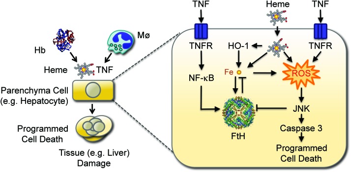
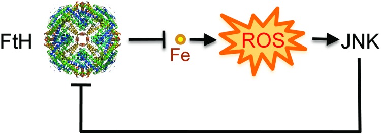
Recent advances: Heme release from hemoglobin sensitizes parenchyma cells to undergo programmed cell death in response to proinflammatory cytokines, such as tumor necrosis factor. This cytotoxic effect is driven by a mechanism involving intracellular accumulation of free radicals, which sustain the activation of the c-Jun N-terminal kinase (JNK) signaling transduction pathway. While heme catabolism by heme oxygenase-1 (HO-1) prevents programmed cell death, this cytoprotective effect requires the co-expression of ferritin H (heart/heavy) chain (FTH), which controls the pro-oxidant effect of labile Fe released from the protoporphyrin IX ring of heme. This antioxidant effect of FTH restrains JNK activation, whereas JNK activation inhibits FTH expression, a cross talk that controls metabolic adaptation to cellular Fe overload associated with systemic infections.

Critical issues and future directions: Identification and characterization of the mechanisms via which FTH provides metabolic adaptation to tissue Fe overload should provide valuable information to our current understanding of the pathogenesis of systemic infections as well as other immune-mediated inflammatory diseases.


Figures

<b>FIG. 1.</b>
FIG. 1.
Cellular Fe homeostasis. Fe metabolism is maintained at a cellular level by a series of evolutionary conserved mechanisms regulating intracellular Fe uptake, trafficking, and export. The scheme provides an overlook of these mechanisms without taking into account species and/or cell specificities. Extracellular Fe (yellow circles) exists in plasma mainly bound to transferrin (TF). Fe uptake occurs via a mechanism that involves the recognition of Fe-TF complexes by transferrin receptor 1 (TFR1), transferrin receptor 2 (TFR2), or Cubilin. Upon internalization of Fe-TF complexes by endocytosis, the acidification of the endolysosomal compartment allows for Fe release. Labile Fe is reduced and transported into the cytoplasm by divalent metal transporter 1 (DMT1). In the specific context of Fe absorption by enterocytes, DMT1 is used to absorb Fe from the intestinal lumen. Intracellular labile Fe is directed mainly to the mitochondria, being imported by Fe transporters, which include the mitochondrial Fe importer mitoferrin-2 (Mfrn2). In the mitochondria, Fe is used for heme biosynthesis and Fe-sulfur cluster assembly or stored by mitochondrial ferritin (not depicted in the scheme). When intracellular labile Fe accumulates above a certain threshold level, Fe can be stored and neutralized intracellularly by multimeric ferritin complexes composed of FTH and FTL chains or exported by the Fe transporters ferroportin (FPN). Fe, iron; FTH, ferritin H (heart/heavy) chain; FTL, ferritin liver/light chain. To see this illustration in color, the reader is referred to the web version of this article at www.liebertpub.com/ars
<b>FIG. 2.</b>
FIG. 2.
Bioavailable Fe. Only an estimated 20%–25% of the bioavailable Fe in mammals exists in the form of nonheme (labile) Fe, either bound to TF, stored by ferritin, as part of a Fe-Sulfur cluster or transiently bound to Fe chaperones and transporters. The remaining 75%–80% of the bioavailable Fe is contained inside the protoporphyn IX ring of heme as prosthetic groups of hemoproteins. The major hemoproteins compartments in mammals are hemoglobin (Hb) in red blood cells (RBCs), myoglobin in muscle cells, and cytochromes in virtual all cell types. Hb contains an estimated 70% of the total pool of heme, myoglobin 5%–10%, and cytochromes 25%–20%, respectively. Fe can transit from the “nonheme” to the “heme” compartment through de novo heme synthesis, a process driven by a sequence of eight enzymatic steps, the last being catalyzed by ferrochelatase, which inserts Fe inside the proptoporphyrin IX ring giving rise to heme. Fe can also transit from the heme to the nonheme pool via the catabolism of heme by HO enzymes or via heme oxidation, which releases Fe from the proptoporphyrin IX ring of heme. HO, heme oxygenases. To see this illustration in color, the reader is referred to the web version of this article at www.liebertpub.com/ars
<b>FIG. 3.</b>
FIG. 3.
Cellular heme homeostasis. When released from Hb, nonprotein bound heme, that is, free heme, is captured in plasma by hemopexin (HPX) or by albumin (Alb), forming heme-hemopexin and heme-Alb complexes, respectively. Cell-free Hb can also be captured in plasma by haptoglobin (HP), forming Hb-haptoglobin (HP) complexes, from which heme release is inhibited. Plasma Hb-haptoglobin and heme-hemopexin complexes are recognized by the macrophage transmembrane CD163 and the low-density lipoprotein receptor-related protein 1 (CD91) receptors, respectively, and internalized into endolysosomes. Possibly, extracellular heme-Alb or eventually “free heme” can also be internalized via the heme transporters feline leukemia virus C receptor 2 (FLVCR2), the heme responsive gene-1 (HRG-1), and the heme carrier protein 1 (HCP1), although this remains to be formally established. Intracellular heme transits from endolysosomes to the cytoplasm via a mechanism involving HRG-1, giving rise to intracytoplasmic heme, which can be transported into the mitochondria via a heme transporter that remains to be identified (?). A possible involvement of ATP-binding cassette subfamily B member 6 (ABCB6) in heme transfer into mitochondria has been proposed. Mitochondrial heme can be exported to the cytoplasm via the FLVCR1β isoform. Intracytoplasmic heme can be catabolized by HO, a process assisted by FTH, which neutralizes the Fe extracted from the protoporphyrin ring of heme, storing it into multimeric ferritin complexes. Alternatively, intracytoplasmic heme can be exported from cells by the ATP-binding cassette subfamily G member 2 (ABCG2) and the FLVCR1α. Whether heme is transported to the nucleus is likely to be the case, although this has not been established. For a recent and comprehensive review on heme transport, see reference (189). To see this illustration in color, the reader is referred to the web version of this article at www.liebertpub.com/ars
<b>FIG. 4.</b>
FIG. 4.
Systemic Fe homeostasis in mammals. Systemic Fe homeostasis is maintained in mammals through a series of mechanisms regulating Fe absorption, its mobilization, and storage into different compartments [adapted from Hentze et al. (78)]. Considering that daily Fe excretion is minimal and absorption is residual compared to the amount required to sustain erythropoiesis and other vital functions, almost all the Fe used in mammals must be recycled within different compartments of the organism. The main pathways of Fe uptake and trafficking involved in the maintenance of Fe homeostasis at a systemic level are illustrated. Normal values for Fe content in different human tissues are also shown [adapted from Hentze et al. (78)]. Notice that 75%–80% of the bioavailable Fe exists in the form of heme as a prosthetic group of Hb. Fe recycling via phagocytosis of senescent RBCs by hemophagocytic macrophages (Mø) in the red pulp of the spleen insures that Fe is extracted from heme and recycled back via TF to provide a steady-state Fe supply for erythropoiesis in the bone marrow. To see this illustration in color, the reader is referred to the web version of this article at www.liebertpub.com/ars
<b>FIG. 5.</b>
FIG. 5.
Heme release from Hb. Pathogens, products associated with the activation of innate and adaptive immunity, changes in plasma pH and osmolarity, microvascular clotting, vasoconstriction, and molecules released in the context of tissue damage can act directly or indirectly to trigger varying levels of RBC lyses and concomitant Hb leakage into plasma. Upon release from RBC, Hb tetramers are dissociated into dimers favoring oxidation of their prosthetic heme groups and promoting heme release. The shear number of RBC (2–3×10¹³ in humans), their high Hb (3×10⁶ molecules/RBC), and heme (1.2×10⁷ molecules/RBC) content make that lysis of a small fraction of RBC not detectable by standard hematological analyzes can lead to the release of significant amount of heme into plasma. Presumably for this reason, Hb is the main source of free heme involved in the pathogenesis of immune-mediated inflammatory diseases, such as severe sepsis and malaria. To see this illustration in color, the reader is referred to the web version of this article at www.liebertpub.com/ars
<b>FIG. 6.</b>
FIG. 6.
Protective effect of FTH against systemic infection. Disruption of RBCs produces cell-free Hb, that upon oxidation releases heme. Non-Hb (free) heme sensitizes hepatocytes to undergo programmed cell death in response to TNF. This cytotoxic effect is mediated via a mechanism involving the production of free radicals (reactive oxygen species; ROS) that sustain JNK activation, leading to caspase activation and ultimately to programmed cell death by apoptosis. Labile Fe released from the protoporphyrin ring of heme catalyzes the production of free radicals via the Fenton chemistry, sustaining JNK activation and leading to programmed cell death. Labile Fe, however, also induces FTH expression, which neutralizes its pro-oxidant effects, suppressing JNK activation and programmed cell death. Expression of FTH can also be induced via NF-κB activation in response to TNF. Expression of FTH is inhibited by JNK activation, promoting the accumulation of labile Fe and the production of ROS, leading to programmed cell death. This pathological process can compromise host survival when confronted with systemic infections, as demonstrated for severe forms of malaria. JNK, c-Jun N-terminal kinase; NF-κB, nuclear factor kappa B; TNF, tumor necrosis factor. To see this illustration in color, the reader is referred to the web version of this article at www.liebertpub.com/ars
<b>FIG. 7.</b>
FIG. 7.
Induction of heme catabolism by sickle cell trait confers disease tolerance to malaria. Sickle cell disease is a molecular disease caused by a single-point mutation in the β chain of Hb (β6Glu>Val). When present in the heterozygous form, the Hb β6Glu>Val sickle mutation is not pathogenic, conferring a survival advantage against malaria (sickle cell trait). This protective effect acts via the accumulation of low (noncytotoxic) levels of free heme in plasma that induce the expression of HO-1 via a mechanism involving the activation of the transcription factor NF-E2-related factor 2 (NRF2) (not illustrated). The CO produced via heme catabolism by HO-1 binds to cell-free Hb and prevents the accumulation of free heme following Plasmodium infection, thus suppressing the pathogenesis of severe forms of malaria. This protective effect does not interfere with parasite load revealing that sickle Hb confers disease tolerance to malaria. CO, carbon monoxide. To see this illustration in color, the reader is referred to the web version of this article at www.liebertpub.com/ars
<b>FIG. 8.</b>
FIG. 8.
FTH prevents pro-oxidant labile Fe from sustaining JNK activation. FTH controls JNK activation indirectly via a mechanism that prevents labile iron from partaking in the production of free radicals via the Fenton chemistry. This is consistent with previous studies showing that reducing free radical production is sufficient per se to control JNK activity, via inhibition of redox-sensitive phosphatases regulating JNK activity (not illustrated). Presumably, the mechanism underlying the cytotoxic effect of JNK activation involves the inhibition of FTH expression, which promotes cellular Fe overload, accumulation of free radicals, and programmed cell death. This functional cross talk between FTH and JNK controls the host metabolic adaptation to tissue Fe overload during systemic infections. To see this illustration in color, the reader is referred to the web version of this article at www.liebertpub.com/ars

References

    1. Abergel RJ, Wilson MK, Arceneaux JE, Hoette TM, Strong RK, Byers BR, and Raymond KN. Anthrax pathogen evades the mammalian immune system through stealth siderophore production. Proc Natl Acad Sci U S A 103: 18499–18503, 2006 - PMC - PubMed
    1. Ackerman D. and Gems D. Insulin/IGF-1 and hypoxia signaling act in concert to regulate iron homeostasis in Caenorhabditis elegans. PLoS Genet 8: e1002498, 2012 - PMC - PubMed
    1. Aisen P, Leibman A, and Zweier J. Stoichiometric and site characteristics of the binding of iron to human transferrin. J Biol Chem 253: 1930–1937, 1978 - PubMed
    1. Ajioka RS, Phillips JD, and Kushner JP. Biosynthesis of heme in mammals. Biochim Biophys Acta 1763: 723–736, 2006 - PubMed
    1. Alam J, Stewart D, Touchard C, Boinapally S, Choi AM, and Cook JL. NRF2, a Cap'n'Collar transcription factor, regulates induction of the heme oxygenase-1 gene. J Biol Chem 274: 26071–26078, 1999 - PubMed

Publication types