

Impact of in vivo reflectance confocal microscopy on the number needed to treat melanoma in doubtful lesions
- PMID: 24124911
- PMCID: PMC3984366
- DOI: 10.1111/bjd.12678
Impact of in vivo reflectance confocal microscopy on the number needed to treat melanoma in doubtful lesions
Abstract
Background: The number needed to treat (NNT) ratio is an effective method for measuring accuracy in melanoma detection. Dermoscopy reduces the number of false positives and subsequently unnecessary excisions. In vivo reflectance confocal microscopy (RCM) is a noninvasive technique that allows examination of the skin with cellular resolution.
Objectives: To assess the impact of RCM analysis on the number of equivocal lesions, assumed to be melanocytic, excised for every melanoma.
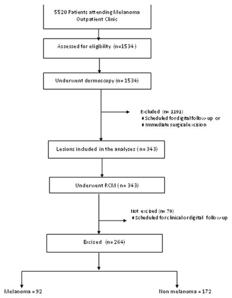
Methods: Consecutive patients (n = 343) presenting with doubtful lesions were considered for enrolment. The lesions were analysed by dermoscopy and RCM, with histopathological assessment considered the reference standard. The main outcome was the NNT, calculated as the proportion of equivocal lesions excised for every melanoma.
Results: Dermoscopy alone obtained a hypothetical NNT of 3·73; the combination of dermoscopy and RCM identified 264 equivocal lesions that qualified for excision, 92 of which were confirmed to be a melanoma, resulting in an NNT of 2·87, whereas the analysis of RCM images classified 103 lesions as melanoma, with a consequent NNT of 1·12. The difference in the reduction of this ratio was statistically significant between the three groups (P < 0·0001). There was no significant improvement in sensitivity when comparing the combination of dermoscopy and RCM with RCM alone (94·6% vs. 97·8%; P = 0·043). However, the differences between specificities were statistically significant (P < 1 × 10(-6) ), favouring RCM alone.
Conclusion: The addition of RCM analysis to dermoscopy reduces unnecessary excisions with a high diagnostic accuracy and could be a means for reducing the economic impact associated with the management of skin cancer.
Keywords: confocal microscopy; dermoscopy; melanoma; number needed to treat.
© 2013 British Association of Dermatologists.
Conflict of interest statement
Figures
Similar articles
-
Role of In Vivo Reflectance Confocal Microscopy in the Analysis of Melanocytic Lesions.Acta Dermatovenerol Croat. 2018 Apr;26(1):64-67. Acta Dermatovenerol Croat. 2018. PMID: 29782304 Review.
-
Reflectance confocal microscopy for diagnosing cutaneous melanoma in adults.Cochrane Database Syst Rev. 2018 Dec 4;12(12):CD013190. doi: 10.1002/14651858.CD013190. Cochrane Database Syst Rev. 2018. PMID: 30521681 Free PMC article.
-
In vivo reflectance confocal microscopy of equivocal melanocytic lesions detected by digital dermoscopy follow-up.J Eur Acad Dermatol Venereol. 2015 Oct;29(10):1918-25. doi: 10.1111/jdv.13067. Epub 2015 Mar 6. J Eur Acad Dermatol Venereol. 2015. PMID: 25752663
-
Three-Dimensional Total Body Photography, Digital Dermoscopy, and in vivo Reflectance Confocal Microscopy for Follow-Up Assessments of High-Risk Patients for Melanoma: A Prospective, Controlled Study.Dermatology. 2024;240(5-6):803-813. doi: 10.1159/000541894. Epub 2024 Oct 8. Dermatology. 2024. PMID: 39378859 Free PMC article.
-
Reflectance confocal microscopy as a second-level examination in skin oncology improves diagnostic accuracy and saves unnecessary excisions: a longitudinal prospective study.Br J Dermatol. 2014 Nov;171(5):1044-51. doi: 10.1111/bjd.13148. Epub 2014 Oct 19. Br J Dermatol. 2014. PMID: 24891083
Cited by
-
Improving clinical diagnosis of early-stage cutaneous melanoma based on Raman spectroscopy.Br J Cancer. 2018 Nov;119(11):1339-1346. doi: 10.1038/s41416-018-0257-9. Epub 2018 Nov 9. Br J Cancer. 2018. PMID: 30410059 Free PMC article.
-
One Step Melanoma Surgery for Patient with Thick Primary Melanomas: "To Break the Rules, You Must First Master Them!".Open Access Maced J Med Sci. 2018 Feb 9;6(2):367-371. doi: 10.3889/oamjms.2018.084. eCollection 2018 Feb 15. Open Access Maced J Med Sci. 2018. PMID: 29531606 Free PMC article.
-
"Twin lesions": Which one is the bad one? Improvement of clinical diagnosis with reflectance confocal microscopy.Dermatol Pract Concept. 2017 Jan 31;7(1):11-17. doi: 10.5826/dpc.0701a02. eCollection 2017 Jan. Dermatol Pract Concept. 2017. PMID: 28243488 Free PMC article.
-
In vivo Study to Evaluate an Intelligent Algorithm for Time Efficient Detection of Malignant Melanoma Using Dermatofluoroscopy.Skin Pharmacol Physiol. 2024;37(4-6):97-103. doi: 10.1159/000542854. Epub 2024 Dec 2. Skin Pharmacol Physiol. 2024. PMID: 39622207 Free PMC article.
-
Reflectance confocal microscopy of skin in vivo: From bench to bedside.Lasers Surg Med. 2017 Jan;49(1):7-19. doi: 10.1002/lsm.22600. Epub 2016 Oct 27. Lasers Surg Med. 2017. PMID: 27785781 Free PMC article. Review.
References
-
- Balch CM, Soong S-J, Atkins MB, et al. An evidence-based staging system for cutaneous melanoma. CA Cancer J Clin. 2004;54:131–49. - PubMed
-
- Balch CM, Soong SJ, Gershenwald JE, et al. Prognostic factors analysis of 17,600 melanoma patients: validation of the American Joint Committee on Cancer melanoma staging system. J Clin Oncol. 2001;19:3622–34. - PubMed
-
- Vestergaard ME, Macaskill P, Holt PE, Menzies SW. Dermoscopy compared with naked eye examination for the diagnosis of primary melanoma: a meta-analysis of studies performed in a clinical setting. Br J Dermatol. 2008;159:669–76. - PubMed
-
- Kittler H, Pehamberger H, Wolff K, Binder M. Diagnostic accuracy of dermoscopy. Lancet Oncol. 2002;3:159–65. - PubMed
Publication types
MeSH terms
Grants and funding
LinkOut - more resources
Full Text Sources
Other Literature Sources
Medical
