. 2013 Oct 29;110(44):17969-74.
doi: 10.1073/pnas.1309362110. Epub 2013 Oct 14.

ASPP2 suppresses squamous cell carcinoma via RelA/p65-mediated repression of p63

Affiliations

ASPP2 suppresses squamous cell carcinoma via RelA/p65-mediated repression of p63

Luca Tordella et al. Proc Natl Acad Sci U S A. .

Abstract

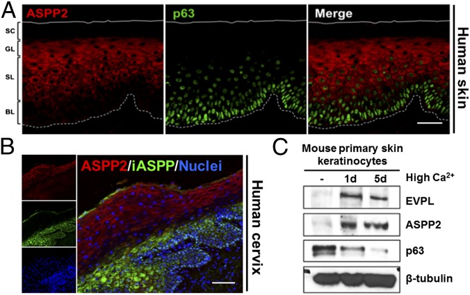
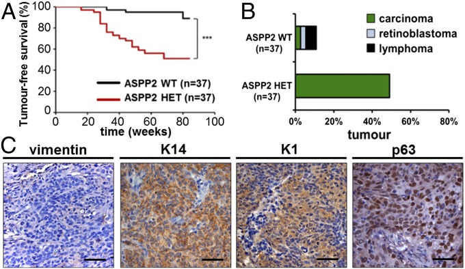
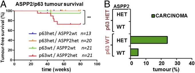
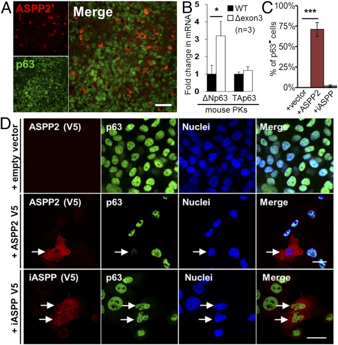
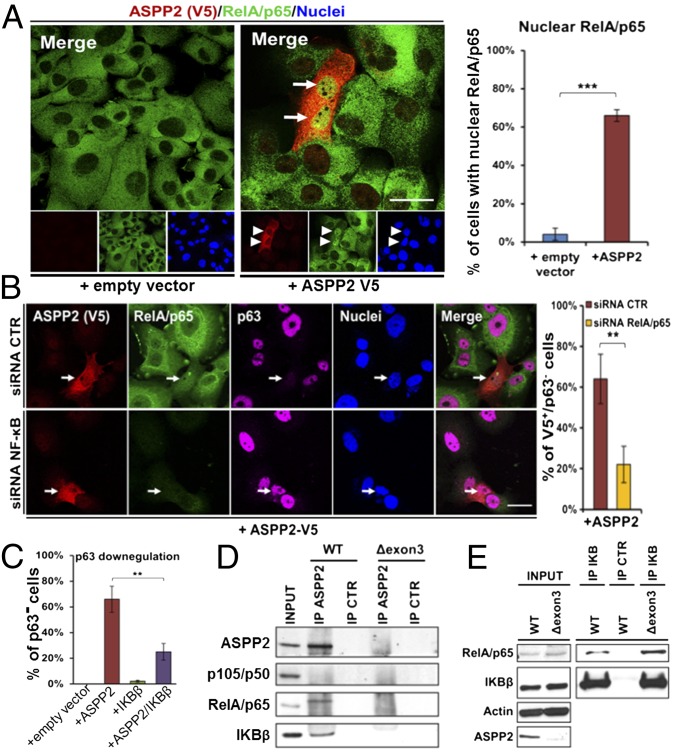
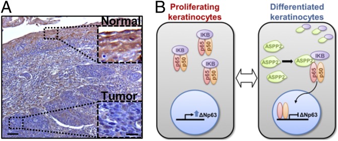
Squamous cell carcinoma (SCC) is highly malignant and refractory to therapy. The majority of existing mouse SCC models involve multiple gene mutations. Very few mouse models of spontaneous SCC have been generated by a single gene deletion. Here we report a haploinsufficient SCC mouse model in which exon 3 of the Tp53BP2 gene (a p53 binding protein) was deleted in one allele in a BALB/c genetic background. Tp53BP2 encodes ASPP2 (ankyrin repeats, SH3 domain and protein rich region containing protein 2). Keratinocyte differentiation induces ASPP2 and its expression is inversely correlated with p63 protein in vitro and in vivo. Up-regulation of p63 expression is required for ASPP2(Δexon3/+) BALB/c mice to develop SCC, as heterozygosity of p63 but not p53 prevents them from developing it. Mechanistically, ASPP2 inhibits ΔNp63 expression through its ability to bind IκB and enhance nuclear Rel/A p65, a component of the NF-κB transcription complex, which mediates the repression of p63. Reduced ASPP2 expression associates with tumor metastasis and increased p63 expression in human head and neck SCCs. This study identifies ASPP2 as a tumor suppressor that suppresses SCC via inflammatory signaling through NF-κB-mediated repression of p63.

Keywords: T53BP2; inflammation; stratified epithelial cell tumor.


Conflict of interest statement

The authors declare no conflict of interest.

Figures

Fig. 1.
Fig. 1.
Mutually exclusive expression of ASPP2 and p63 in squamous epithelia and PKs. (A) Double staining of human skin squamous epithelium using anti-ASPP2 and anti-p63 antibodies. BL, basal layer; GL, granular layer; SC, stratum corneum; SL, spinous layer. (B) ASPP2 and iASPP double IF staining of human cervical epithelium sections. Nuclei were counterstained with DAPI. (C) Immunoblot shows expression levels of ASPP2, p63, and envoplakin (EVPL) in Ca2+-induced mouse PK with β-tubulin as loading control. (Scale bars: 50 µm.)
Fig. 2.
Fig. 2.
ASPP2Δexon3/+ BALB/c mice develop spontaneous SCC with high frequency and early onset. (A) Tumor-free survival and (B) tumor incidence and spectrum spontaneously developed in ASPP2+/+ (WT) and ASPP2Δexon3/+ (HET) BALB/c mice over 88 wk [***P = 0.0002, log-rank (Mantel–Cox) test]. (C) Immunohistochemical staining of vimentin, K14, K1, and p63 in tumor sections. (Scale bars: 50 µm.)
Fig. 3.
Fig. 3.
Heterozygosity of p63 prevents ASPP2Δexon3/+ BALB/c mice from developing SCC. (A) Tumor-free survival and (B) tumor incidence and spectrum in ASPP2/p63 compound mice with indicated genotypes over 80 wk [**P = 0.0086 by log-rank (Mantel–Cox) test].
Fig. 4.
Fig. 4.
ASPP2 represses p63 expression in vivo and in vitro. (A) Double IF staining of ASPP2 and p63 in a tumor section derived from an ASPP2Δexon3/+ BALB/c mouse. (Scale bar: 20 µm.) (B) RT-qPCR expression analysis of ΔNp63 and TAp63 mRNA in mouse PKs (whole epidermis), with GAPDH mRNA as internal control. (*P = 0.034; n indicates the number of littermate-paired PKs used). Bar graph values are the mean ± SD from three different experiments. (C and D) Double IF staining of 040 cells to detect transfected ASPP2-V5 (red) or iASPP-V5 (red) and endogenous p63 (green). TO-PRO was used to visualize nuclei. ASPP2-V5+ and iASPP-V5+ cells are labeled with white arrows (D). The percentage of V5+/p63 cells in indicated transfected samples is shown in C (***P < 0.0001). Values are mean ± SD from three different experiments. (Scale bars: 10 µm.)
Fig. 5.
Fig. 5.
ASPP2 inhibits p63 by inducing nuclear p65/NFκB. (A) Double IF staining of 040 cells using anti-V5 (red) and anti-RelA/p65 (green) antibodies. Arrows label cells expressing ASPP2-V5 and nuclear RelA/p65. The graph shows the percentage of nuclear RelA/p65 expressing cells in indicated transfected samples (***P < 0.001). (B) Triple IF staining of 040 cells to detect transfected ASPP2-V5 (red), endogenous RelA/p65 (green), and endogenous p63 (magenta) in cells treated with control (CTR) or RelA/p65 (NFκB) RNAi. Arrows label ASPP2-V5–expressing cells. The bar graph shows the percentage of V5+/p63 cells in transfected samples as indicated. (**P = 0.0083). (C) Graph shows the percentage of cells with low or undetectable p63 in transfected samples as indicated. (**P = 0.001). (D and E) Lysates from ASPP2 WT and Δexon3 MEFs were immunoprecipitated with a control IgG (IP CTR) or indicated antibodies (IP ASPP2, IP IκB) and immunoblotted with antibodies as labeled. (Scale bars: 10 µm.) Bar graph values are the mean ± SD from three different experiments.
Fig. 6.
Fig. 6.
ASPP2 expression is decreased in human SCC samples. (A) ASPP2 immunostain of human HNSCC. Areas of normal epithelium and tumor mass are shown at higher magnification. (Scale bars: 50 µm and 10 µm.) (B) Diagram to summarize the interplay between ASPP2, IκB, and RelA/p65 in regulating ΔNp63 expression.

References

    1. Quintanilla M, Brown K, Ramsden M, Balmain A. Carcinogen-specific mutation and amplification of Ha-ras during mouse skin carcinogenesis. Nature. 1986;322(6074):78–80. - PubMed
    1. Vitale-Cross L, Amornphimoltham P, Fisher G, Molinolo AA, Gutkind JS. Conditional expression of K-ras in an epithelial compartment that includes the stem cells is sufficient to promote squamous cell carcinogenesis. Cancer Res. 2004;64(24):8804–8807. - PubMed
    1. Kemp CJ, Donehower LA, Bradley A, Balmain A. Reduction of p53 gene dosage does not increase initiation or promotion but enhances malignant progression of chemically induced skin tumors. Cell. 1993;74(5):813–822. - PubMed
    1. Martínez-Cruz AB, et al. Spontaneous squamous cell carcinoma induced by the somatic inactivation of retinoblastoma and Trp53 tumor suppressors. Cancer Res. 2008;68(3):683–692. - PubMed
    1. Qiao W, et al. Hair follicle defects and squamous cell carcinoma formation in Smad4 conditional knockout mouse skin. Oncogene. 2006;25(2):207–217. - PubMed

Publication types

MeSH terms

LinkOut - more resources