. 2013 Nov 26;7(11):9761-70.
doi: 10.1021/nn403258w. Epub 2013 Oct 17.

Gold nanocrystal labeling allows low-density lipoprotein imaging from the subcellular to macroscopic level

Affiliations

Gold nanocrystal labeling allows low-density lipoprotein imaging from the subcellular to macroscopic level

Iris E Allijn et al. ACS Nano. .

Abstract

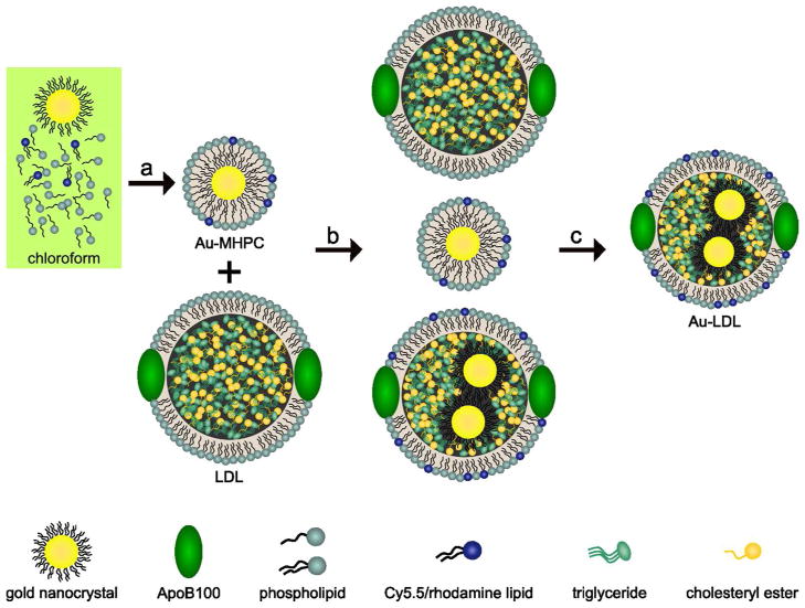
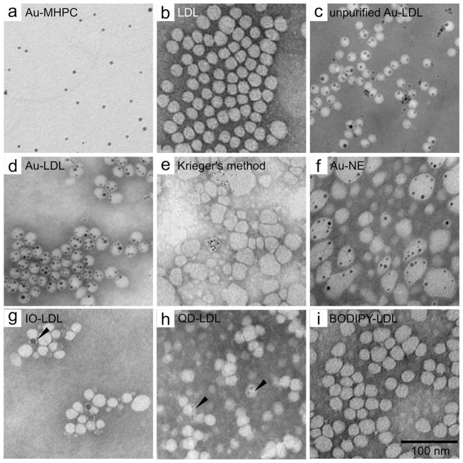
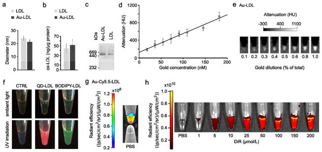
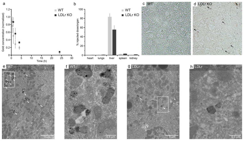
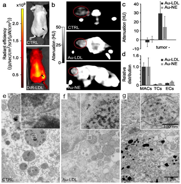
Low-density lipoprotein (LDL) plays a critical role in cholesterol transport and is closely linked to the progression of several diseases. This motivates the development of methods to study LDL behavior from the microscopic to whole-body level. We have developed an approach to efficiently load LDL with a range of diagnostically active nanocrystals or hydrophobic agents. We performed focused experiments on LDL labeled with gold nanocrystals (Au-LDL). The labeling procedure had minimal effect on LDL size, morphology, or composition. Biological function was found to be maintained from both in vitro and in vivo experiments. Tumor-bearing mice were injected intravenously with LDL, DiR-LDL, Au-LDL, or a gold-loaded nanoemulsion. LDL accumulation in the tumors was detected with whole-body imaging methods, such as computed tomography (CT), spectral CT, and fluorescence imaging. Cellular localization was studied with transmission electron microscopy and fluorescence techniques. This LDL labeling procedure should permit the study of lipoprotein biointeractions in unprecedented detail.


Figures

Figure 1
Figure 1. Labeling schematic of low density lipoprotein
Dodecanethiol coated gold nanocrystals were mixed with phospholipids and Cy5.5 lipids in CHCL3. a, This solution was dripped into >70 °C H2O, forming micelles enclosing a gold nanocrystal. b, The phospholipid coated gold nanocrystals were mixed with native LDL in PBS and sonicated yielding three different particles: free phospholipid-coated gold nanocrystals, native LDL, and native LDL with gold nanocrystals in the hydrophobic core. c, This solution was purified on a dual density gradient, resulting in a solution of gold labeled LDL nanoparticles (Au-LDL).
Figure 2
Figure 2. LDL labeled with different payloads
Negative stain TEM micrographs of: a, phospholipid-coated gold nanocrystals; b, native human LDL; c, unpurified Au-LDL; d, purified Au-LDL; e, TEM of LDL labeled with 3 nm Au nanocrystals using Krieger’s reconstitution method. Most LDL nanoparticles stay unlabeled and their morphology has changed. f, TEM of Au-NE nanoemulsions, the control particle used in the animal experiments. g, TEM of 10 nm IO-LDL. Most LDL stays unlabeled, some cores seem to attach to the side of the LDL, but not enter the core (arrowhead). h, TEM of QD-LDL. QDs are visible in the core of the LDL (arrowheads). i, TEM of BODIPY-LDL. Scale bar is the same for all TEM micrographs. NE = nano emulsion, IO = iron oxide, QD = quantum dot.
Figure 3
Figure 3. Characterization of labeled LDL
a, Diameters of LDL and Au-LDL derived from TEM. b, ELISA on oxidation of LDL and Au-LDL. c, Western blot of ApoB100 of Au-LDL and LDL. d, Gold concentration – attenuation curve of Au-LDL phantom. e, Phantom of increasing Au-LDL concentrations. f, Photograph of CTRL, QD-LDL, and BODIPY-LDL in ambient light and under UV irradiation. g, Phantom of Au-Cy5.5-LDL compared with control PBS. h, Fluorescence images of DiR-LDL.
Figure 4
Figure 4. In vitro competition inhibition assay
Fluorescence microscopy of LDL (control) (a–c), Au-LDL (test) (d–f) and LDL + Au-LDL (inhibition competition) (g–h) in HepG2, J774A.1 and B16-F10 cells. CT of control and Au-LDL in HepG2 (j), J774A.1 (k) and B16-F10 (l) cells. Red fluorescence is Au-LDL, blue fluorescence is DAPI.
Figure 5
Figure 5. In vivo functionality of Au-LDL in wild type and LDLr KO mice
a, Pharmacokinetics of Au-LDL in mice. b, Biodistribution of Au-LDL in mice at 2 hr post-injection. c,d, Light microscopy of sliver stained sections of mouse livers. Black arrowheads indicate intense areas of silver staining. e–h, TEM of hepatocytes in mouse livers. White arrowheads indicate accumulations of gold nanoparticles. f and h are higher magnification images of the boxed areas in e and g, respectively.
Figure 6
Figure 6. In vivo and ex vivo imaging
a, IVIS of mice, arrowheads indicate the B16-F10 tumor. b, CT of mice, the tumors are circled. c, Tumor attenuation relative to control. Attenuation in the tumors of the Au-LDL mice were higher than in the Au-NE mice (p=0.009). d, FACS of tumor tissue. MACs: macrophages, TCs; tumor cells, ECs: endothelial cells. e, TEM of control tumor tissue, inset shows no gold. f, TEM of Au-LDL tumor tissue, inset shows gold particles in a vesicle. g, TEM of Au-NE tumor tissue, inset shows gold particles in a vesicle.
Figure 7
Figure 7. Spectral CT of Au-LDL injected B16-F10 tumor bearing mice
Gold accumulation is displayed in yellow and overlaid on conventional CT images. a, 3D reconstruction of the rear of a mouse injected with Au-LDL. Gold accumulation can be observed in the tumor. b, 3D reconstruction of the front of a mouse with gold accumulation found in the liver.

Comment in

References

    1. Corbin IR, Li H, Chen J, Lund-Katz S, Zhou R, Glickson JD, Zheng G. Low-Density Lipoprotein Nanoparticles as Magnetic Resonance Imaging Contrast Agents. Neoplasia. 2006;8:488–498. - PMC - PubMed
    1. Brown M, Goldstein J. Receptor-Mediated Endocytosis: Insights from the Lipoprotein Receptor System. Proc Natl Acad Sci USA. 1979;76:3330–3337. - PMC - PubMed
    1. Segrest J, Jones M, De Loof H, Dashti N. Structure of Apolipoprotein B-100 in Low Density Lipoproteins. J Lipid Res. 2001;42:1346–1367. - PubMed
    1. Firestone RA. Low-Density Lipoprotein as a Vehicle for Targeting Antitumor Compounds to Cancer Cells. Bioconjugate Chem. 1994;5:105–113. - PubMed
    1. Berliner J, Heinecke J. The Role of Oxidized Lipoproteins in Atherogenesis. Free Radic Biol Med. 1996;20:707–727. - PubMed

Publication types