

Sporadic naturally occurring melanoma in dogs as a preclinical model for human melanoma
- PMID: 24128326
- PMCID: PMC4066658
- DOI: 10.1111/pcmr.12185
Sporadic naturally occurring melanoma in dogs as a preclinical model for human melanoma
Abstract
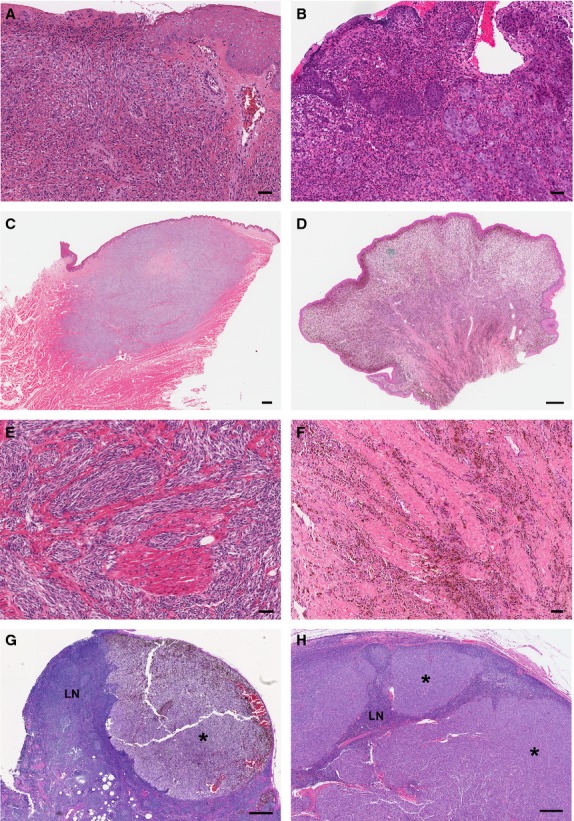
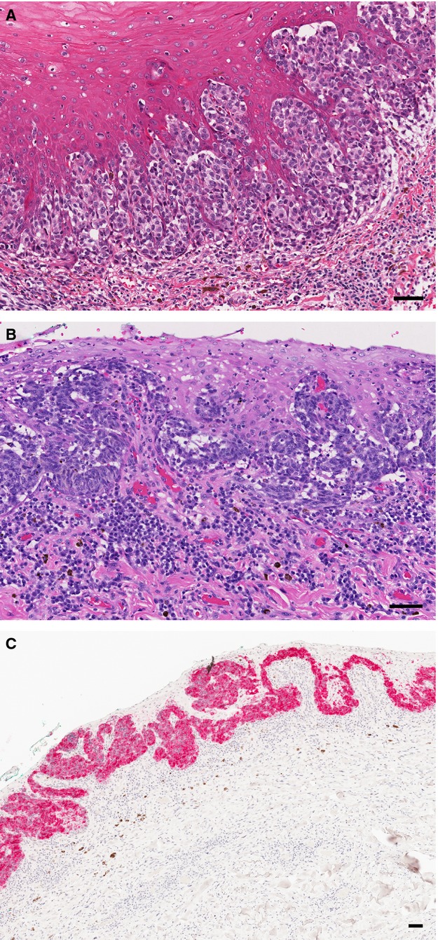
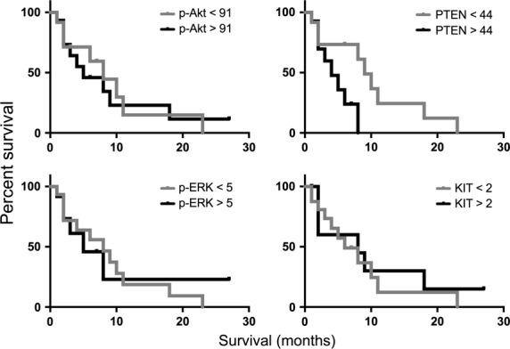
Melanoma represents a significant malignancy in humans and dogs. Different from genetically engineered models, sporadic canine melanocytic neoplasms share several characteristics with human disease that could make dogs a more relevant preclinical model. Canine melanomas rarely arise in sun-exposed sites. Most occur in the oral cavity, with a subset having intra-epithelial malignant melanocytes mimicking the in situ component of human mucosal melanoma. The spectrum of canine melanocytic neoplasia includes benign lesions with some analogy to nevi, as well as invasive primary melanoma, and widespread metastasis. Growing evidence of distinct subtypes in humans, differing in somatic and predisposing germ-line genetic alterations, cell of origin, epidemiology, relationship to ultraviolet radiation and progression from benign to malignant tumors, may also exist in dogs. Canine and human mucosal melanomas appear to harbor BRAF, NRAS, and c-kit mutations uncommonly, compared with human cutaneous melanomas, although both species share AKT and MAPK signaling activation. We conclude that there is significant overlap in the clinical and histopathological features of canine and human mucosal melanomas. This represents opportunity to explore canine oral cavity melanoma as a preclinical model.
Keywords: animal model; clinical trial design; comparative study; digital telepathology; image analysis; melanoma; signal transduction.
© 2013 The Authors. Pigment Cell & Melanoma Research published by John Wiley & Sons Ltd.
Figures
Comment in
-
Melanoma in mankind's best friend.Pigment Cell Melanoma Res. 2014 Jan;27(1):1. doi: 10.1111/pcmr.12196. Pigment Cell Melanoma Res. 2014. PMID: 24344626 No abstract available.
References
-
- Beadling C, Jacobson-Dunlop E, Hodi FS. KIT gene mutations and copy number in melanoma subtypes. Clin. Cancer Res. 2008;14:6821–6828. - PubMed
-
- Bergman P. Wolchok J. Of Mice and Men (and Dogs): development of a xenogeneic DNA vaccine for canine oral malignant melanoma. Cancer Ther. 2008;6:817–826.
-
- Bergman PJ, Kent MS. Farese JP. Melanoma. In: Page RL, editor; Withrow SJ, Vail DM, editors. Withrow and MacEwen's Small Animal Clinical Oncology. St. Louis, MO: Elsevier/Saunders; 2013. pp. 321–334.
-
- Bogenrieder T. Herlyn M. The molecular pathology of cutaneous melanoma. Cancer Biomark. 2010;9:267–286. - PubMed
Publication types
MeSH terms
Substances
Grants and funding
LinkOut - more resources
Full Text Sources
Other Literature Sources
Medical
Research Materials
Miscellaneous
