Review
. 2014 Jan;184(1):18-27.
doi: 10.1016/j.ajpath.2013.08.029. Epub 2013 Oct 12.

LDL receptor-related protein-1: a regulator of inflammation in atherosclerosis, cancer, and injury to the nervous system

Affiliations
Review

LDL receptor-related protein-1: a regulator of inflammation in atherosclerosis, cancer, and injury to the nervous system

Steven L Gonias et al. Am J Pathol. 2014 Jan.

Abstract

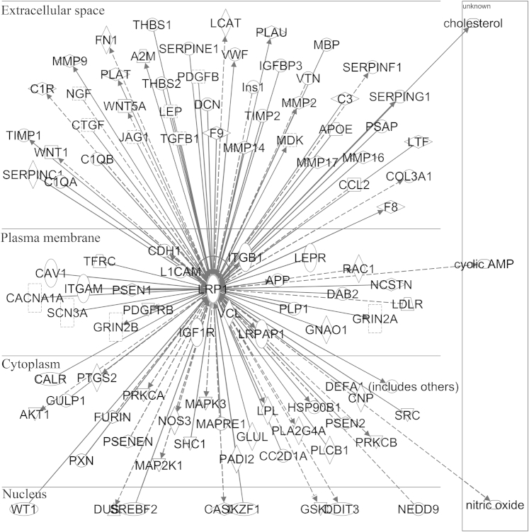
Low-density lipoprotein receptor-related protein-1 (LRP1) is an endocytic receptor for numerous proteins that are both structurally and functionally diverse. In some cell types, LRP1-mediated endocytosis is coupled to activation of cell signaling. LRP1 also regulates the composition of the plasma membrane and may, thereby, indirectly regulate the activity of other cell-signaling receptors. Given the scope of LRP1 ligands and its multifunctional nature, it is not surprising that numerous biological activities have been attributed to this receptor. LRP1 gene deletion is embryonic-lethal in mice. However, elegant studies using Cre-LoxP recombination have helped elucidate the function of LRP1 in mature normal and pathological tissues. One major theme that has emerged is the role of LRP1 as a regulator of inflammation. In this review, we will describe evidence for LRP1 as a regulator of inflammation in atherosclerosis, cancer, and injury to the nervous system.


Figures

Figure 1
Figure 1
Molecular models showing the organization of structural domains in LRP1 and the docking of a representative ligand to complement-like repeats in LRP1. A: The depicted domains in LRP1 are common to the LDL receptor family. Stars are present in the intracellular region of LRP1 to represent motifs that function as endocytosis signals and/or as docking sites for cell-signaling proteins including NPXY, YxxL, and dileucine. B: A representative LRP1 ligand, the 18-kDa receptor-binding domain of α2-macroglobulin, which is a free-standing domain in the activated state of the protein, is shown in pink. Two lysine residues in a single α-helix, highlighted in blue, are essential for binding to LRP1. These lysine residues interact with acidic amino acids in the LRP1 complement-like repeats. The fourth and fifth complement-like repeats in CCR2 are shown in orange, and the acidic amino acids in these domains are highlighted in black. The approximate positions of calcium are shown. EGF, epidermal growth factor.
Figure 2
Figure 2
LRP1 interaction map generated using the Ingenuity IPA System. The map was limited to interactions involving the nervous system in inflammatory disease and in the inflammatory response. Interactions are stratified in relation to the location of the LRP1-interacting gene product relative to the cell. Interacting gene products include cytokines (closed square), growth factors (broken square), proteases (horizontally elongated diamond), other enzymes (vertically elongated diamond), proteins involved in transport (trapezoid), transmembrane receptors (vertically elongated oval), ion channel subunits (broken vertically elongated rectangle), kinases (inverted triangle), transcription regulators (horizontally elongated oval), and phosphatases (triangle). Other categories of gene products are shown as circles. Direct interactions are shown with a solid line and indirect interactions with a broken line. A solid line without an arrow implies a binding interaction. An arrowhead at the end of a broken line implies that one gene product acts on the other. A perpendicular bar implies an inhibitory interaction.
Figure 3
Figure 3
IHC analysis to detect LRP1 in the nervous system. A: LRP1 immunostaining of mouse cerebral cortex. LRP1 was detected with a primary polyclonal antibody that detects the β-chain (Sigma-Aldrich, St. Louis, MO). Staining was conducted using a Ventana System (Tucson, AZ). B: A section of rat sciatic nerve distal to a crush injury site, which was recovered 3 days after nerve injury. LRP1 was detected using polyclonal antibody R2629. The section is counterstained with methylene green. Representative LRP1-immunopositive Schwann cells are marked with arrows. The asterisk marks a macrophage. Scale bars: 35 μm (A); 12 μm (B).

References

    1. Herz J., Hamann U., Rogne S., Myklebost O., Gausepohl H., Stanley K.K. Surface location and high affinity for calcium of a 500-kd liver membrane protein closely related to the LDL-receptor suggest a physiological role as lipoprotein receptor. EMBO J. 1988;7:4119–4127. - PMC - PubMed
    1. Herz J., Kowal R.C., Goldstein J.L., Brown M.S. Proteolytic processing of the 600 kd low density lipoprotein receptor-related protein (LRP) occurs in a trans-Golgi compartment. EMBO J. 1990;9:1769–1776. - PMC - PubMed
    1. Van Leuven F., Cassiman J.J., Van Den Berghe H. Primary amines inhibit recycling of alpha 2M receptors in fibroblasts. Cell. 1980;20:37–43. - PubMed
    1. Herz J., Kowal R.C., Ho Y.K., Brown M.S., Goldstein J.L. Low density lipoprotein receptor-related protein mediates endocytosis of monoclonal antibodies in cultured cells and rabbit liver. J Biol Chem. 1990;265:21355–21362. - PubMed
    1. Wu L., Gonias S.L. The low-density lipoprotein receptor-related protein-1 associates transiently with lipid rafts. J Cell Biochem. 2005;96:1021–1033. - PubMed

Publication types

MeSH terms

Substances