

Maxillary neurilemmoma-Rarest of the rare tumour: Report of 2 cases
- PMID: 24129122
- PMCID: PMC3825964
- DOI: 10.1016/j.ijscr.2013.09.006
Maxillary neurilemmoma-Rarest of the rare tumour: Report of 2 cases
Abstract
Introduction: Intraoral neurilemmomas (schwannoma) are rare, even rarer are intraosseous ones, and the rarest are the maxillary neurilemmomas. Going by the literature only 5 cases of maxillary neurilemomas are reported till now. Neurilemmomas are benign tumours of nerve sheath origin. Approximately 30% arise in head and neck region, of these 1-12% arise intraorally mainly involving tongue.
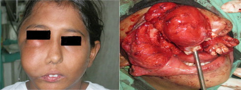
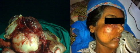
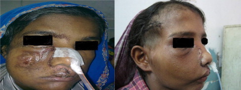
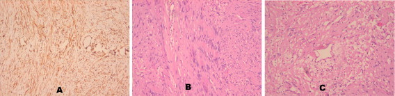
Presentation of case: Here we report two cases of maxillary neurilemmoma, one in a 9 year old girl and second one in a 27 year old female, both involving the lateral surface of maxilla on right side. Both the patients presented with a long standing history of swelling which was increasing gradually. 9-Year-old girl also had 1 lesion in the temporal region on right side and the 27-year-old patient had associated erosion of the soft palate. Diagnosis was made on the basis of histopathology and immunohistochemistry.
Discussion: Neurilemmomas are slow growing benign tumour of the nerve sheath origin arising from the Schwann cells. Their aetiology is not known. Most common complaint is that of a gradually increasing swelling followed by pain and paresthesias. Surgery remains the treatment of choice with close follow up.
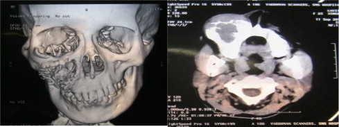
Conclusion: Maxillary neurilemmomas are rarest of the rare tumour which closely mimic benign odontogenic cysts and tumours, and should be kept in the differential diagnosis of these lesions. Knowledge of the radiologic and clinical behaviour of these tumours is extremely important for prompt diagnosis and treatment.
Keywords: Intraoral; Intraosseous; Maxillary; Neurilemmoma; Schwannoma.
Copyright © 2013 The Authors. Published by Elsevier Ltd.. All rights reserved.
Figures
References
-
- Batsakis J.G. 2nd edition. Williams and Wilkins; Baltimore: 1979. Tumours of the peripheral nervous system; pp. 313–333.
-
- Buric N., Jovanovich G., Pesic Z., Krasic D., Radovanovic Z., Mihailovic D. Mandible schwannoma (neurilemmoma) presenting as apical lesion. Dentomaxillofac Radiol. 2009;38:178–181. - PubMed
-
- Colreavy M.P., Lacy P.D., Hughes J., Bouchier-Hayes D., Brennan P., O’Dwyer A.J. Head and neck schwannomas – a 10 year review. J Laryngo Otol. 2000;114:119–124. - PubMed
-
- Chi A.C., Carey J., Muller S. Intraosseous schwannoma of the mandible: a case report and review of the literature. Oral Surg Oral Med Oral Pathol Oral Radiol Endod. 2003;96(1):54–65. - PubMed
-
- Fawcett K.J., Dahlin D.C. Neurilemmoma of bone. Am J Clin Pathol. 1967;47(6):759–766. - PubMed
LinkOut - more resources
Full Text Sources
Other Literature Sources
Research Materials
