

Inhibition of melanogenesis by gallic acid: possible involvement of the PI3K/Akt, MEK/ERK and Wnt/β-catenin signaling pathways in B16F10 cells
- PMID: 24129178
- PMCID: PMC3821624
- DOI: 10.3390/ijms141020443
Inhibition of melanogenesis by gallic acid: possible involvement of the PI3K/Akt, MEK/ERK and Wnt/β-catenin signaling pathways in B16F10 cells
Abstract
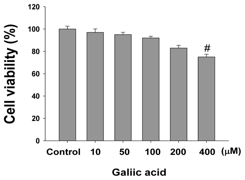
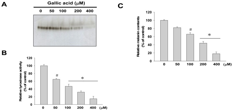
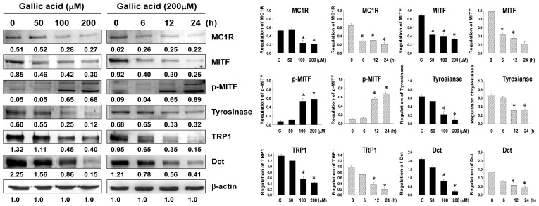
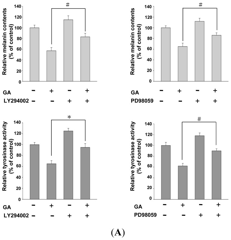
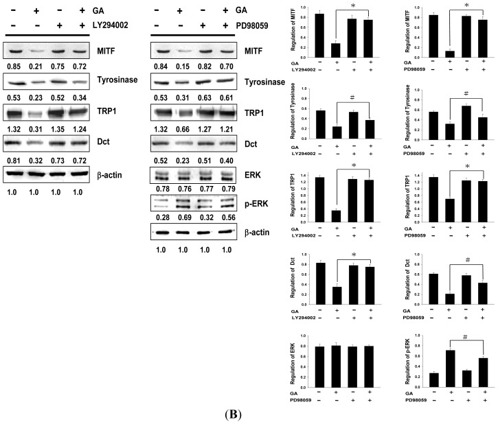
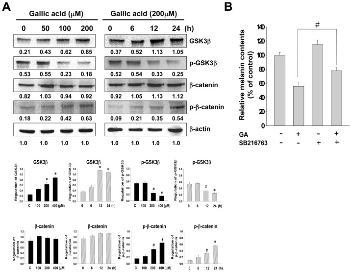
Gallic acid is one of the major flavonoids found in plants. It acts as an antioxidant, and seems to have anti-inflammatory, anti-viral, and anti-cancer properties. In this study, we investigated the effects of gallic acid on melanogenesis, including the activation of melanogenesis signaling pathways. Gallic acid significantly inhibited both melanin synthesis and tyrosinase activity in a dose- and time-dependent manner, and decreased the expression of melanogenesis-related proteins, such as microphthalmia-associated transcription factor (MITF), tyrosinase, tyrosinase-related protein-1 (TRP1), and dopachrome tautomerase (Dct). In addition, gallic acid also acts by phosphorylating and activating melanogenesis inhibitory proteins such as Akt and mitogen-activated protein kinase (MEK)/extracellular signal-regulated kinase (ERK). Using inhibitors against PI3K/Akt (LY294002) or MEK/ERK-specific (PD98059), the hypopigmentation effect was suppressed, and the gallic acid-initiated activation of MEK/ERK and PI3K/Akt was also revoked. Gallic acid also increased GSK3β and p-β-catenin expression but down-regulated p-GSK3β. Moreover, GSK3β-specific inhibitor (SB216763) restored gallic acid-induced melanin reduction. These results suggest that activation of the MEK/ERK, PI3K/Akt, and inhibition of Wnt/β-catenin signaling pathways is involved in the melanogenesis signaling cascade, and that activation by gallic acid reduces melanin synthesis via down-regulation of MITF and its downstream signaling pathway. In conclusion, gallic acid may be a potentially agent for the treatment of certain skin conditions.
Figures

Similar articles
-
Partially purified components of Nardostachys chinensis suppress melanin synthesis through ERK and Akt signaling pathway with cAMP down-regulation in B16F10 cells.J Ethnopharmacol. 2011 Oct 11;137(3):1207-14. doi: 10.1016/j.jep.2011.07.047. Epub 2011 Jul 26. J Ethnopharmacol. 2011. PMID: 21816215
-
Betulinic acid isolated from Vitis amurensis root inhibits 3-isobutyl-1-methylxanthine induced melanogenesis via the regulation of MEK/ERK and PI3K/Akt pathways in B16F10 cells.Food Chem Toxicol. 2014 Jun;68:38-43. doi: 10.1016/j.fct.2014.03.001. Epub 2014 Mar 12. Food Chem Toxicol. 2014. PMID: 24632067
-
Pyruvic acid/ethyl pyruvate inhibits melanogenesis in B16F10 melanoma cells through PI3K/AKT, GSK3β, and ROS-ERK signaling pathways.Genes Cells. 2019 Jan;24(1):60-69. doi: 10.1111/gtc.12654. Epub 2018 Dec 18. Genes Cells. 2019. PMID: 30417494
-
The Hypopigmentation Mechanism of Tyrosinase Inhibitory Peptides Derived from Food Proteins: An Overview.Molecules. 2022 Apr 22;27(9):2710. doi: 10.3390/molecules27092710. Molecules. 2022. PMID: 35566061 Free PMC article. Review.
-
The Modulation of Melanogenesis in B16 Cells Upon Treatment with Plant Extracts and Isolated Plant Compounds.Molecules. 2022 Jul 7;27(14):4360. doi: 10.3390/molecules27144360. Molecules. 2022. PMID: 35889231 Free PMC article. Review.
Cited by
-
Attenuation of melanogenesis by Nymphaea nouchali (Burm. f) flower extract through the regulation of cAMP/CREB/MAPKs/MITF and proteasomal degradation of tyrosinase.Sci Rep. 2018 Sep 17;8(1):13928. doi: 10.1038/s41598-018-32303-7. Sci Rep. 2018. PMID: 30224716 Free PMC article.
-
Grape Extract Promoted α-MSH-Induced Melanogenesis in B16F10 Melanoma Cells, Which Was Inverse to Resveratrol.Molecules. 2021 Oct 1;26(19):5959. doi: 10.3390/molecules26195959. Molecules. 2021. PMID: 34641503 Free PMC article.
-
Fermented Aronia melanocarpa Inhibits Melanogenesis through Dual Mechanisms of the PI3K/AKT/GSK-3β and PKA/CREB Pathways.Molecules. 2023 Mar 27;28(7):2981. doi: 10.3390/molecules28072981. Molecules. 2023. PMID: 37049743 Free PMC article.
-
Wnt3a promotes radioresistance via autophagy in squamous cell carcinoma of the head and neck.J Cell Mol Med. 2019 Jul;23(7):4711-4722. doi: 10.1111/jcmm.14394. Epub 2019 May 21. J Cell Mol Med. 2019. PMID: 31111621 Free PMC article.
-
Gomisin N Inhibits Melanogenesis through Regulating the PI3K/Akt and MAPK/ERK Signaling Pathways in Melanocytes.Int J Mol Sci. 2017 Feb 22;18(2):471. doi: 10.3390/ijms18020471. Int J Mol Sci. 2017. PMID: 28241436 Free PMC article.
References
-
- Tsatmali M., Ancans J., Thody A.J. Melanocyte function and its control by melanocortin peptides. J. Histochem. Cytochem. 2002;50:125–133. - PubMed
-
- Briganti S., Camera E., Picardo M. Chemical and instrumental approaches to treat hyperpigmentation. Pigment Cell Melanoma Res. 2003;16:101–110. - PubMed
-
- Busca R., Ballotti R. Cyclic AMP a key messenger in the regulation of skin pigmentation. Pigment Cell Res. 2000;13:60–69. - PubMed
Publication types
MeSH terms
Substances
LinkOut - more resources
Full Text Sources
Other Literature Sources
Research Materials
Miscellaneous
