. 2013 Oct 15;4(5):e00737-13.
doi: 10.1128/mBio.00737-13.

Origin, evolution, and genotyping of emergent porcine epidemic diarrhea virus strains in the United States

Affiliations

Origin, evolution, and genotyping of emergent porcine epidemic diarrhea virus strains in the United States

Yao-Wei Huang et al. mBio. .

Abstract

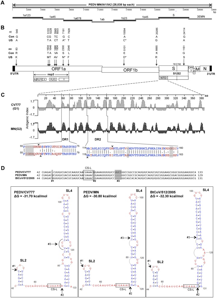
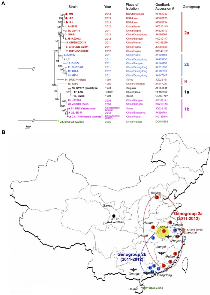
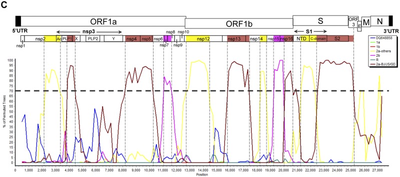
Coronaviruses are known to infect humans and other animals and cause respiratory and gastrointestinal diseases. Here we report the emergence of porcine epidemic diarrhea virus (PEDV) in the United States and determination of its origin, evolution, and genotypes based on temporal and geographical evidence. Histological lesions in small intestine sections of affected pigs and the complete genomic sequences of three emergent strains of PEDV isolated from outbreaks in Minnesota and Iowa were characterized. Genetic and phylogenetic analyses of the three U.S. strains revealed a close relationship with Chinese PEDV strains and their likely Chinese origin. The U.S. PEDV strains underwent evolutionary divergence, which can be classified into two sublineages. The three emergent U.S. strains are most closely related to a strain isolated in 2012 from Anhui Province in China, which might be the result of multiple recombination events between different genetic lineages or sublineages of PEDV. Molecular clock analysis of the divergent time based on the complete genomic sequences is consistent with the actual time difference, approximately 2 to 3 years, of the PED outbreaks between China (December 2010) and the United States (May 2013). The finding that the emergent U.S. PEDV strains share unique genetic features at the 5'-untranslated region with a bat coronavirus provided further support of the evolutionary origin of PEDV from bats and potential cross-species transmission. The data from this study have important implications for understanding the ongoing PEDV outbreaks in the United States and will guide future efforts to develop effective preventive and control measures against PEDV.

Importance: The sudden emergence of porcine epidemic diarrhea virus (PEDV), a coronavirus, for the first time in the United States causes significant economic and public health concerns. Since its recognition in May 2013, PEDV has rapidly spread across the United States, resulting in high mortality in piglets in more than 17 States now. The ongoing outbreaks of Middle East respiratory syndrome coronavirus in humans from countries in or near the Arabian Peninsula and the historical deadly nature of the 2002 outbreaks of severe acute respiratory syndrome coronavirus create further anxiety over the emergence of PEDV in the United States due to the lack of scientific information about the origin and evolution of this emerging coronavirus. Here we report the detailed genetic characterization, origin, and evolution of emergent PEDV strains in the United States. The results provide much needed information to devise effective preventive and control strategies against PEDV in the United States.


Figures

FIG 1
FIG 1
Schematic diagrams of genomic structure, the strategy for genomic cDNA cloning, and molecular characterization of unique features of three emergent U.S. PEDV strains (MN, IA1, and IA2) isolated in Minnesota and Iowa in 2013. (A) Eight overlapping cDNA fragments covering the entire PEDV genome for each of the three U.S. strains, represented by thick lines, were amplified by RT-PCR from total RNAs extracted from fecal or small intestine samples. The names of each fragment are indicated. The location and primer sequences for the RT-PCR and the RACE-PCR used to generate the overlapping RT-PCR products are available in Table S1 in the supplemental material. The numbers on the scale bar indicate distances from the 5′ end of the genome. (B) Organization of the PEDV genome and locations of unique amino acid (aa) changes identified in the three U.S. strains. The approximate positions and sizes of genes in the PEDV genome that correspond to the scale bar are shown in panel A. The putative S1/S2 boundary (amino acid positions) of the S protein is also shown. Nucleotide (nt) and amino acid differences between the 3 emergent U.S. PEDV sequences and the consensus sequences of 23 other known PEDV strains and their positions are depicted. Con, PEDV consensus sequences; US, unique sequences in three U.S. PEDV strains; *, unique amino acids and nucleotides shared by the three U.S. strains and one Chinese AH2012 strain; UTR, untranslated region; nsp, nonstructural protein; Ac, acidic domain; PLP, papain-like proteinase; X, X domain (ADP-ribose-1′-phosphatase [ADRP]); Y, unknown Y domain; NTD, N-terminal domain of the spike gene. (C) Comparison of antigenic index profiles of the NTD (aa 1 to 380) of S protein between the prototype strain CV777 (genogroup 1 [G1]) and U.S. strain MN (genogroup 2 [G2]). The corresponding alignment of amino acid sequences and positions of the two regions containing amino acid deletions/insertions (indicated by dashes and shaded) are shown. DR, region containing deletions. Identical amino acids are marked in blue, whereas mismatches are marked in red. Favorable amino acid mismatches are displayed as colons, whereas neutral mismatches are depicted as periods. The predicted N-linked glycosylation sites in DR1 and DR2 are indicated by arrowheads. (D) Comparisons of the primary sequences and the predicted secondary structures of a 5′-proximal region in the 5′-UTR between Belgian PEDV/CV777 (nt 42 to 133), U.S. PEDV/MN (nt 42 to 129), and a bat coronavirus BtCoV/512/2005 (nt 44 to 131). Three deletions of nucleotides (no. 1 to 3) are indicated by dashes and shaded in the primary sequences and are indicated by arrows in the secondary structures. The core sequences (CUAAAC) of leader transcription-regulating sequences (CS-L) are boxed by dashed lines. Two stem-loops (SL2 and SL4) conserved in all coronaviruses are indicated. (There is no SL3 in alphacoronaviruses.) Nucleotides in the stems are marked in blue, whereas nucleotides in the loops are marked in red.
FIG 2
FIG 2
(Continued)
FIG 2
FIG 2
Genotyping and origin of the emergent U.S. PEDV strains based on full-length genomic sequence analyses. (A) Phylogeny-based genotyping of 26 PEDV strains with available complete genomic sequences, including the 3 U.S. PEDV strains. The tree was constructed by the neighbor-joining method, based upon the full-length genomic nucleotide sequences using the bat coronavirus BtCoV/512/2005 sequence as an outgroup. Bootstrap values are indicated for each node from 1,000 resamplings. The names of the strains, years and places of isolation, GenBank accession numbers, and genogroups and subgroups proposed in this study are shown. (An asterisk indicates that the isolation year of LZC is unknown but should be before 2006 according to the GenBank submission date.) Red solid circles, the three U.S. PEDV strains; purple solid triangles, cell-culture-adapted PEDV strains or vaccines; green solid diamonds, bat coronavirus BtCoV/512/2005. (B) Phylogeny-based geographical dissection of genogroup 2 Chinese PEDV strains. The map of China shows all of the provinces where genogroup 2 PEDV strains with the available complete genomic sequences were isolated. The numbers in order and the colors for PEDV strains correspond to those labeled in panel A: genogroup 2a strains are in red, and genogroup 2b strains are in blue. The coverage area for each subgroup (depicted by the red or the blue oval) is deduced based on the distributions of the strains. XS indicates a representative strain (XS2012) isolated in Zhejiang Province of eastern China that belongs to genogroup 2a based on the sequence of the S gene from this study. The yellow shaded circle indicates the hypothetical location of the origin of the U.S. PEDV, where the closely related AH2012 strain was identified. The two early PEDV strains CH/S and LZC and their locations are also shown. Five provinces where bat coronaviruses phylogenetically related to PEDV were isolated (10) are also marked by the “bat” symbols. In particular, the BtCoV/512/2005 strain, isolated in Hainan Province, is marked in green. (C) Bootscan analysis for possible recombination events of lineage US-AH strains (three U.S. strains plus the AH2012 strain) in subgroup 2a as the query group throughout the genome compared to the other two lineages. 2a-BJ/JS/GD includes strains BJ-2011-1, GD-B and JS-HZ2012 (denoted by the brown line), and “2a-others” includes CH/ZMDZY/11, CH/FJND-3/2011, and CH/FLZZ-9/2012 (denoted by the yellow line) in the same subgroup (based upon the phylogenetic tree constructed in panel A), subgroup 1a (green line), 1b (red line), R (cyan line), 2b (purple line), and BtCoV/512/2005 (accession no. DQ648858 [blue line]). Bootscanning was conducted by SimPlot (version 3.5.1; window size: 1,000 bp; step, 200 bp), and the cutoff value of bootstrap support for clustering was set to 70. The putative recombinant regions corresponding to functional domains in the PEDV genome are shown at the top.

Similar articles

Cited by

References

    1. de Groot RJ, Baker SC, Baric R, Enjuanes L, Gorbalenya AE, Holmes KV, Perlman S, Poon L, Rottier PJM, Talbot PJ, Woo PCY, Ziebuhr J. 2011. Coronaviridae, p 806–828 In King AMQ, Adams MJ, Carstens EB, Lefkowitz EJ. (ed), Virus taxonomy: ninth report of the International Committee on Taxonomy of Viruses. Elsevier Academic, London, United Kingdom
    1. Brian DA, Baric RS. 2005. Coronavirus genome structure and replication. Curr. Top. Microbiol. Immunol. 287:1–30 - PMC - PubMed
    1. Woo PC, Lau SK, Lam CS, Lau CC, Tsang AK, Lau JH, Bai R, Teng JL, Tsang CC, Wang M, Zheng BJ, Chan KH, Yuen KY. 2012. Discovery of seven novel mammalian and avian coronaviruses in the genus Deltacoronavirus supports bat coronaviruses as the gene source of Alphacoronavirus and Betacoronavirus and avian coronaviruses as the gene source of Gammacoronavirus and Deltacoronavirus. J. Virol. 86:3995–4008 - PMC - PubMed
    1. Graham RL, Baric RS. 2010. Recombination, reservoirs, and the modular spike: mechanisms of coronavirus cross-species transmission. J. Virol. 84:3134–3146 - PMC - PubMed
    1. van Boheemen S, de Graaf M, Lauber C, Bestebroer TM, Raj VS, Zaki AM, Osterhaus AD, Haagmans BL, Gorbalenya AE, Snijder EJ, Fouchier RA. 2012. Genomic characterization of a newly discovered coronavirus associated with acute respiratory distress syndrome in humans. mBio 3(6):e00473-12.10.1128/mBio.00473-12 - DOI - PMC - PubMed

MeSH terms