. 2013 Oct 15;4(5):e00745-13.
doi: 10.1128/mBio.00745-13.

Streptococcus pneumoniae biofilm formation is strain dependent, multifactorial, and associated with reduced invasiveness and immunoreactivity during colonization

Affiliations

Streptococcus pneumoniae biofilm formation is strain dependent, multifactorial, and associated with reduced invasiveness and immunoreactivity during colonization

Krystle Blanchette-Cain et al. mBio. .

Abstract

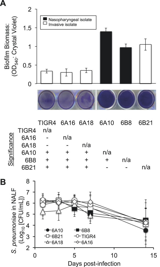
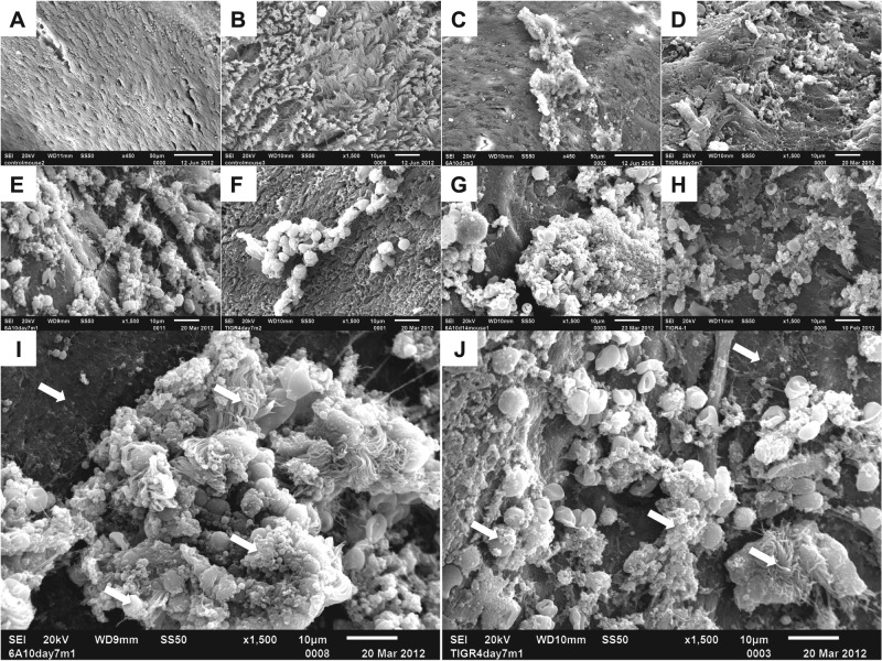
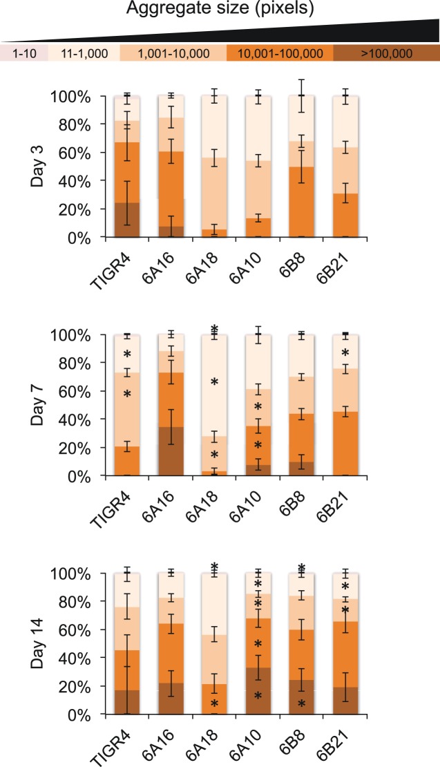
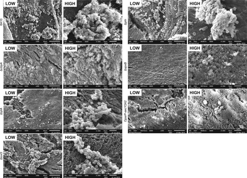
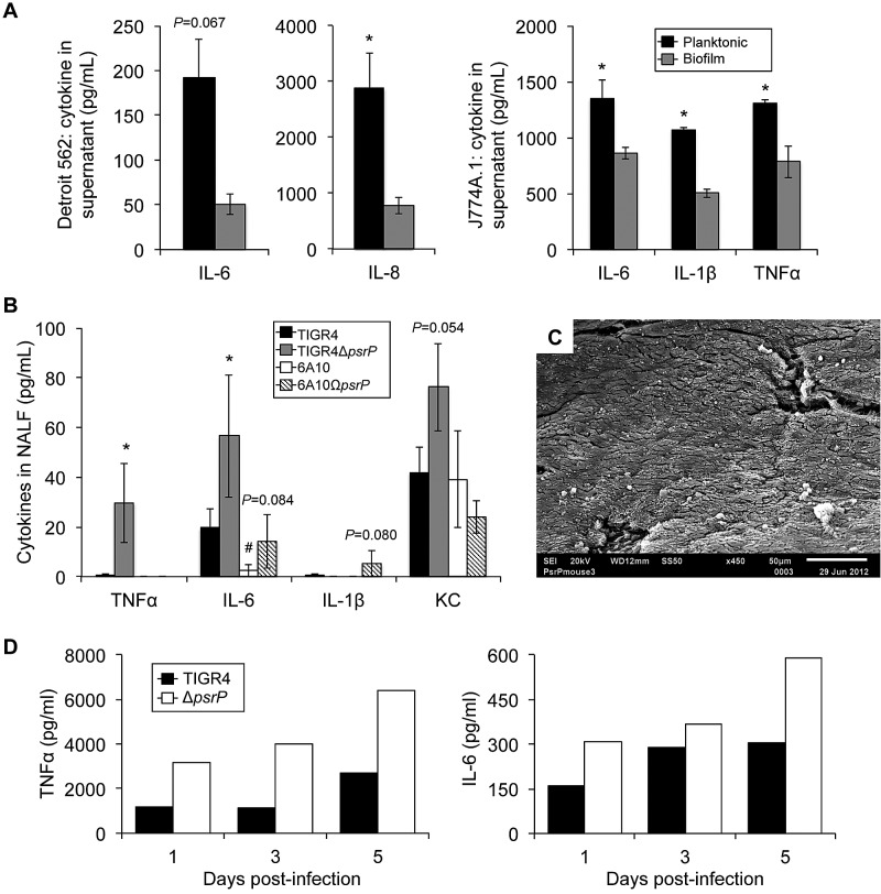
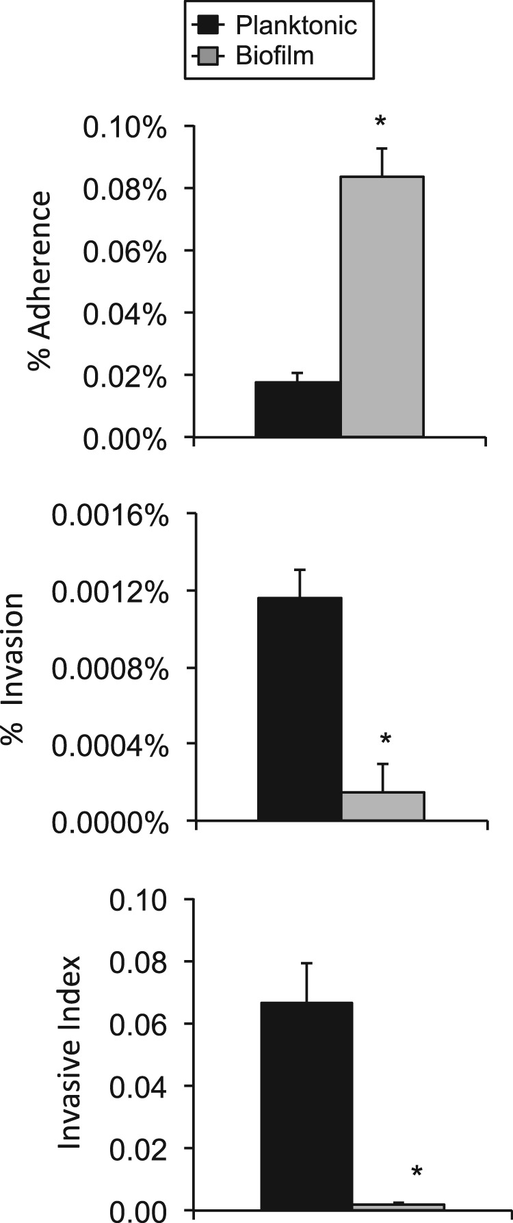
Biofilms are thought to play an important role during colonization of the nasopharynx by Streptococcus pneumoniae, yet how they form in vivo and the determinants responsible remain unknown. Using scanning electron microscopy, we show that biofilm aggregates of increasing complexity form on murine nasal septa following intranasal inoculation. These biofilms were highly distinct from in vitro biofilms, as they were discontiguous and appeared to incorporate nonbacterial components such as intact host cells. Biofilms initially formed on the surface of ciliated epithelial cells and, as cells were sloughed off, were found on the basement membrane. The size and number of biofilm aggregates within nasal lavage fluid were digitally quantitated and revealed strain-specific capabilities that loosely correlated with the ability to form robust in vitro biofilms. We tested the ability of isogenic mutants deficient in CbpA, pneumolysin, hydrogen peroxide, LytA, LuxS, CiaR/H, and PsrP to form biofilms within the nasopharynx. This analysis revealed that CiaR/H was absolutely required for colonization, that PsrP and SpxB strongly impacted aggregate formation, and that other determinants affected aggregate morphology in a modest fashion. We determined that mice colonized with ΔpsrP mutants had greater levels of the proinflammatory cytokines tumor necrosis factor alpha (TNF-α), interleukin-6 (IL-6), IL-1β, and KC in nasal lavage fluid than did mice colonized with wild-type controls. This phenotype correlated with a diminished capacity of biofilm pneumococci to invade host cells in vitro despite enhanced attachment. Our results show that biofilms form during colonization and suggest that they may contribute to persistence through a hyperadhesive, noninvasive state that elicits a dampened cytokine response.

Importance: This work demonstrates the first temporal characterization of Streptococcus pneumoniae biofilm formation in vivo. Our results show that the morphology of biofilms formed by both invasive and noninvasive clinical isolates in vivo is distinct from that of formed biofilms in vitro, yet propensity to form biofilms in vivo loosely correlates with the degree of in vitro biofilm formation on a microtiter plate. We show that host components, including intact host cells, influence the formation of in vivo structures. We also found that efficient biofilm formation in vivo requires multiple bacterial determinants. While some factors are essential for in vivo biofilm formation (CiaRH, PsrP, and SpxB), other factors are less critical (CbpA, LytA, LuxS, and pneumolysin). In comparison to their planktonic counterparts, biofilm pneumococci are hyperadhesive but less invasive and elicit a weaker proinflammatory cytokine response. These findings give insight into the requirements for and potential role of biofilms during prolonged asymptomatic colonization.


Figures

FIG 1
FIG 1
In vitro biofilm formation by S. pneumoniae clinical isolates and corresponding rates of colonization. (A) Mean biomass of biofilms formed by clinical isolates on 6-well polystyrene plates after 18 h of growth, as measured by crystal violet staining (n = 4/strain). Accompanying representative images of stained wells are provided beneath the graph. The table represents statistical significance between strains (+, significance; −, no significance) grown on untreated plates as determined by one-way ANOVA. n/a, not applicable; OD540, optical density at 540 nm. (B) CFU determination of S. pneumoniae in NALF collected from colonized mice at days 1, 3, 5, 7, and 14 postinfection. No significant differences were observed between strains on any given day as tested by one-way ANOVA (n = 9 to 12/cohort).
FIG 2
FIG 2
Pneumococcal biofilms are present on mucosal epithelial cells in the septa of experimentally infected mice. Scanning electron microscopy of isolated nasal septa from naive or colonized mice. (A and B) Low (A)- and high (B)-magnification views of healthy septal epithelia. (C) 6A10-colonized nasal septa at 3 dpi. Both matrix-associated bacteria and free diplococci are present on top of ciliated epithelial cells. (D) TIGR4-colonized septa at 3 dpi. Small aggregates of bacteria are present. (E and I) 6A10-colonized septa at 7 dpi. Bacteria are encased within a matrix, and aggregates contain a number of visible host components. Large aggregates are associated with an absence of cilia. (F and J) TIGR4-colonized septa at 7 dpi. Clusters of aggregated bacteria rest on the epithelial surface and are associated with a number of host cells. (G and H) 6A10 (G)- and TIGR4 (H)-colonized respiratory epithelium at 14 dpi. Cilia are absent, and bacterial aggregates are seen on the exposed cells. Bacteria are encased within a thick, visible matrix, and individual cells are difficult to distinguish. 6A10 aggregates are large and highly structured, whereas TIGR4 aggregates were mottled and noticeably smaller. (I and J) Aggregates formed by both 6A10 (I) and TIGR4 (J) contain a number of host components, including inflammatory cells. Arrows indicate common features of biofilm architecture: exposed basement membrane, bacterial cells within matrix material, and incorporated ciliated cells and leukocytes.
FIG 3
FIG 3
In vivo aggregates detected in NALF of mice colonized with S. pneumoniae. Following processing with ImageJ software (see Materials and Methods), individual biofilm aggregates were grouped into classes based on pixel size, from small aggregates (1 to 10/11 to 1,000) to medium (1,001 to 10,000/10,001 to 100,000) to large (>100,000). Shown is the contribution of each size class to the total aggregate area as determined by a percentage of the total pixel area. Asterisks denote a significant difference between size areas compared to day 3. Statistical analysis was done using Student’s t test.
FIG 4
FIG 4
Biofilm aggregates on septal epithelia of mice colonized with isogenic mutants. Nasal septa from 6A10-colonized mice collected at 7 dpi at low and high magnification. Aggregate formation in vivo was minimally decreased or equal to that of the wild type in strains lacking CbpA or pneumolysin, as structures were still fairly large in size and contained host components. Mutants lacking LytA, LuxS, and SpxB exhibited a decreased ability to form large aggregates, and observed aggregates rested on top of ciliated epithelial cells, in contrast to the exposed basement membrane observed in wild-type-colonized samples. The PsrP mutant strain was completely unable to form biofilm aggregates, and colonized mice displayed a healthy ciliated epithelia. The presence of scattered immune cells could be seen.
FIG 5
FIG 5
Mutant strains are able to colonize efficiently but exhibit a decreased ability to form aggregates in vivo. ImageJ quantitation of aggregate size distribution over time by 6A10 or 6A10 isogenic mutants. Statistical significance was determined by 2-tailed Student’s t test versus the 6A10 control at the corresponding time point and is represented with an asterisk.
FIG 6
FIG 6
S. pneumoniae that is unable to form biofilms elicits an enhanced cytokine response during colonization. (A) Assorted cytokine levels in Detroit-562 pharyngeal epithelial cell or J774A.1 macrophage supernatants 24 h following a 4-h exposure to 106 CFU of either planktonic or biofilm-derived TIGR4 pneumococci. (B) Levels of TNF-α, IL-6, IL-1β, and KC in nasal lavage samples collected at 7 days postinfection from mice colonized with wild-type or PsrP-deficient isogenic mutants of strains TIGR4 and 6A10 (n = 5/cohort). Cytokine levels were measured using ELISA. Significance was tested by Student’s t test (n = 5/cohort). Asterisks denote a statistically significant difference (P < 0.05) for the mutant versus its respective wild type. Hash tag denotes statistically significant difference versus TIGR4. (C) Representative scanning electron micrograph of septa isolated from mouse colonized with T4ΔpsrP. Note that T4ΔpsrP does not form the biofilm aggregates observed in mice infected with TIGR4 or cause the sloughing of mucosal epithelial cells. (D) Levels of TNF-α and IL-6 in pooled nasal lavage samples collected from mice colonized with wild-type or PsrP-deficient isogenic mutants of strain TIGR4 on days 1, 3, and 5 postinfection (n = 6/cohort). Cytokine levels were measured using ELISA.
FIG 7
FIG 7
Biofilm bacteria are hyperadhesive but less invasive. Percentage of TIGR4 cells from the total inoculum that attached and invaded Detroit-562 pharyngeal epithelial cells. The ratio of invasive pneumococci to those that are attached is also shown. Experiments were performed in triplicate. Statistical analysis was performed using a two-tailed Student t test. Asterisks denote a significant difference (P < 0.05).

References

    1. Musher D. 2010. Streptococcus pneumoniae, p 2623–2642 In Mandell G, Bennett J, Dolin R. (ed), Mandell, Douglas, and Bennett’s principles and practice of infectious disease, 7th ed, vol 2 Churchill: Livingstone Elsevier, Philadelphia, PA
    1. Reference deleted.
    1. O’Brien KL, Wolfson LJ, Watt JP, Henkle E, Deloria-Knoll M, McCall N, Lee E, Mulholland K, Levine OS, Cherian T, Hib, Pneumococcal Global Burden of Disease Study Team 2009. Burden of disease caused by Streptococcus pneumoniae in children younger than 5 years: global estimates. Lancet 374:893–902 - PubMed
    1. Crook D, Brueggmann A, Sleeman K, Peto T. 2004. Pneumococcal carriage. ASM Press, Washington, DC
    1. Costerton JW, Stewart PS, Greenberg EP. 1999. Bacterial biofilms: a common cause of persistent infections. Science 284:1318–1322 - PubMed

Publication types

Substances