. 2013 Dec;19(12):1711-25.
doi: 10.1261/rna.039461.113. Epub 2013 Oct 15.

Novel small RNA expression libraries uncover hsa-miR-30b and hsa-miR-30c as important factors in anoikis resistance

Novel small RNA expression libraries uncover hsa-miR-30b and hsa-miR-30c as important factors in anoikis resistance

Miguel A Moreno-Mateos et al. RNA. 2013 Dec.

Abstract

MicroRNAs (miRNAs) have been widely studied in order to elucidate their biological functions. MicroRNA microarrays or miRNA overexpression libraries generated by synthesis and cloning of individual miRNAs have been used to study their different roles. In this work, we have developed a novel methodology to express mature miRNAs and other small RNAs from a double convergent RNA polymerase III promoter. We show that the generated miRNAs function similarly to those processed from primary transcripts or pri-miRNAs. This system allowed us to produce a lentiviral library expressing the whole population of small RNAs present in a metastatic cell line. A functional screening using this library led to the identification of hsa-miR-30b and hsa-miR-30c as negative regulators of cell death induced by loss of attachment (anoikis). Importantly, we demonstrated that the acquisition of anoikis resistance via these miRNAs is achieved through down-regulation of caspase 3 expression. Moreover, overexpression of these miRNAs resulted in a decrease of other types of caspase 3-dependent cell death and enhanced the survival of MCF10A acinar cells in morphogenesis assays, suggesting a putative role as oncomirs. In summary, this novel methodology provides a powerful and effective way for identifying novel small RNAs involved in a particular biological process.

Keywords: anoikis; functional screens; libraries; miR-30b/c; small RNAs.


Figures

FIGURE 1.
FIGURE 1.
Mature miRNAs expressed under the control of dual convergent promoters are functionally similar to full-length expressed miRNAs. (A) Schematic drawing showing the plasmid used to express mature miRNAs and others small RNAs. (B) RNase protection assay of miR-21, miR-19a transiently overexpressed from the pLENT-DUAL plasmid in HEK-293T cells (upper and lower panel). miR-21 full-length (miR-21 FL) was expressed from a pCMV-miR-21 plasmid. Control (Ctr) cells expressed the empty vectors pLENT-DUAL or pCMV. Y corresponds to yeast RNA. (C) Transient transfections were carried out in HEK-293T cells, using pLENT-DUAL-21, -19, -15 (expressing the mature form of miR-21, miR-19a or miR-15a, respectively) or pCMV-miR-21 full-length, pLuc-BS carrying a binding site for miR-21, miR-19a or miR-15a and pCMV-Renilla (normalization control) at 100:10:1 proportions, respectively. Firefly and Renilla activities were measured 40 h after transfection. (D) Similar luciferase assay as described in C, using pLENT-DUAL-miR-21 and pCMV-miR-21 full-length and a plasmid harboring a 652- nt wild-type sequence corresponding to the 3′ UTR of PDCD4 at the 3′ position of the luciferase reporter gene. (E) Similar luciferase assay as described in C in HCT116 DICER −/− cells, using pLENT-DUAL-miR-21 and pCMV-miR-21 full-length and a plasmid carrying a binding site for miR-21. For these experiments, HCT116 DICER −/− cells infected with a lentivirus expressing a small hairpin RNA against DGCR8 were used. All transfections were performed in triplicate, and results are shown as the averages ± standard errors of the means from at least three independent experiments. The data were subjected to two-tailed Student's t-test [(*) P < 0.05, (***) P < 0.001], with the exception of section E, where a two-way ANOVA followed by Bonferroni's post-test was used. (*) P < 0.05.
FIGURE 2.
FIGURE 2.
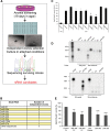
Generation of a small RNA library from MDA-MB-231 (clone 4175) cells. (A) Scheme illustrating the small RNA cloning strategy to generate expression libraries based on the pLENT-DUAL lentiviral vector. (B) Pie chart representing the percentage of the different classes of small RNAs in the pLENT-DUAL library. (Repeats) Regions annotated as repeats (of any type, excluding simple repeats and primarily transposable elements), (scRNA) small cytoplasmic RNA, (other hairpins) loci that look like hairpins but do not pass all the criteria for annotation as miRNAs, (sense strand) small RNA matching sense strand to coding and noncoding regions, (antisense strand) small RNA matching antisense strand to coding and noncoding regions. (C) A correlation between known miRNAs expressed in MDA-MB-231 cells and known miRNAs cloned in the library (log [1 + number of reads normalized to total number of mapped reads]). R2 derived from Pearson's correlation of the data is indicated.
FIGURE 3.
FIGURE 3.
A functional screening using a novel small RNAs expression library reveals hsa-miR-30b and hsa-miR-30c as repressors of anoikis. (A) Immortalized RPE-1 cells were infected with a lentiviral expression library containing a small RNAs collection from the MDA-MB-231 cell line. RPE-1-infected cells were cultured for 19 d in agar dishes at low confluence avoiding any contact and surviving clones transferred to adhesive culture dishes to allow the growth of anoikis-resistant cells. DNA from these clones was isolated and sequenced. (B) Table showing the small RNA sequences overexpressed in anoikis-resistant clones. (C) Flow cytometry analysis of the levels of apoptosis in RPE-1 cells (SubG1/SubG1 control + polyHEMA) infected with lentivirus containing candidate mature miRNAs or empty vector (control). Three days after infection, cells were deprived of serum for 24 h and then cultured in polyhema (PH) plates plus methylcellulose (2%) in serum-free medium for another 24 h. (D) RNase protection assay of miR-30b and miR-30c transiently overexpressed in HEK-293 cells from pLENT-DUAL plasmid. Control (Ctr) cells expressed the empty vector pLENT-DUAL; Y corresponds to yeast RNA; M, Decade RNA markers (Ambion). (E) Levels of apoptosis in RPE-1 cells (SubG1/SubG1 control) infected with lentivirus carrying full-length miR-30b, miR-30c-1 (FL), miR-30b/c mature miRNAs (Lent-30b/c), and the corresponding empty vectors as controls in a similar assay as described in C. Results are shown as the averages ± standard errors of the means from at least three independent experiments and were analyzed by one-way ANOVA, followed by Bonferroni post-test for significance versus control cells. (*) P < 0.05, (**) P < 0.01, (***) P < 0.001.
FIGURE 4.
FIGURE 4.
Hsa-miR-30b and hsa-miR-30c down-regulate CASP3 expression through its 3′ UTR-binding. (A) Western blot analysis of p53, CASP3, and BCL2-like 11 (BIM), three putative target proteins of miR-30b/c related to anoikis. RPE-1 cells were infected with lentivirus expressing full-length miR-30b, miR-30c-1 (miR-30c), or empty vector as a control. Active CASP3 was analyzed in cells cultured under anoikis conditions as described in Figure 3C. Actin (ACTB) was used as the loading control. (B) Densitometric analysis of pro-CASP3 and active CASP3 levels. ACTB protein was used as the normalization control. Results are shown as the averages ± standard errors of the means from at least three independent experiments and were analyzed by one-way ANOVA, followed by Bonferroni post-test (*P < 0.05; **P < 0.01). (C) Schematic representation of the caspase 3 3′ UTR containing two putative binding sites for miR-30b/c. The 3′ UTR of CASP3 mRNA was cloned downstream from the open reading frame of luciferase (pLuc-BS). Both broadly (1187–1193) and poorly (1222–1228) conserved putative miRNA regulatory elements of the CASP3 3′ UTR were mutated (Mut1 and Mut2, respectively) and cloned along with the wild-type 3′ UTR (WT). HEK-293T cells were transiently cotransfected with plasmids overexpressing full-length miRNAs (miR-30b, miR-30c-1, or empty vector as control), pLuc-3′ UTR CASP3 (WT, Mut1, or Mut2) and pCMV-Renilla at 100:10:1 proportions, respectively. Firefly and Renilla activities were measured 40 h after transfection. The luciferase activity normalized to Renilla is shown (lower panel). Transfections were performed in triplicate, and results are shown as the averages ± standard errors of the means from at least three independent experiments and were analyzed by one-way and two-way ANOVA, followed by Bonferroni post-test. (*) P < 0.05, (**) P < 0.01, (***) P < 0.001, (ns) not significant.
FIGURE 5.
FIGURE 5.
Caspase 3 is the main target of miR-30b/c in the anoikis-resistance context. (A) RPE-1 cells were co-infected with both lentiviruses expressing full-length miR-30b/c and the ORF of caspase 3. Western blot analysis of pro-CASP3, active CASP3, and ACTB is shown for RPE-1 cells growing either in complete medium under adherent conditions (−PH) or in anoikis-inducing conditions (+PH). Flow cytometry analysis of the levels of apoptosis (SubG1/SubG1 control) in CASP3-infected cells growing under anoikis-inducing conditions (lower panel). (B) RPE-1 cells were infected with either lentivirus expressing a shRNA to silence the expression of caspase 3 or a scramble shRNA (control). Experiments were carried out with RPE-1 infected cells previously selected in puromycin for 12 d. The diagram to the left shows CASP3 mRNA levels measured by real time PCR after puromycin selection. HPRT mRNA was used as the normalization control. On the right, levels of apoptotic cells in anoikis-inducing conditions are shown. Results are shown as the averages ± standard errors of the means from three independent experiments. The data were subjected to two-tailed Student's t-test. (**) P < 0.01, (***) P < 0.001. (C) Number of colonies after plating 2 × 10⁴ cells from B per well in six-well plates with a bottom layer of 0.5% agar and a top layer of 0.35% agarose. Colonies were photographed and counted after 4 wk. Results are shown as the averages ± standard errors of the means from three independent experiments. The data were subjected to two-tailed Student's t-test. (**) P < 0.01.
FIGURE 6.
FIGURE 6.
Hsa-miR-30a and -30d do not confer anoikis resistance in RPE-1 cells. (A) Western blot analysis of pro-CASP3 in RPE-1 cells infected with lentivirus expressing full-length miR-30a, miR-30d, or their corresponding empty vectors as control. Actin (ACTB) was used as the loading control. On the right, densitometric analysis of pro-CASP3 is shown. ACTB protein was used as a normalization control. (B) Flow cytometry analysis of the levels of apoptosis (SubG1/SubG1 control) in infected cells growing under anoikis-inducing conditions. (C) Endogenous levels of mature miR-30a/d (left panel) and miR-30b/c (right panel) expressed in RPE-1 and MDA-MB-231 (clone 4175) cells. miR-16 and miR-324 were used as normalization controls for miR-30b/c and miR-30a/d, respectively. Similar results were observed when human U6 snRNA primer and U6-snRNA (RNU6B) were used as controls. Results are shown as the averages ± standard errors of the means from three independent experiments. The data were subjected to two-tailed Student's t-test. (*) P < 0.05.
FIGURE 7.
FIGURE 7.
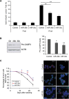
Hsa-miR-30b and hsa-miR-30c reduce cell death induced by TRAIL and delay acini clearance in MCF10A cells. (A) Cells were infected with lentivirus containing full-length miR-30b, miR-30c, and empty vector as a control. Four days after infection, cells were cultured in the presence of Trail (500 ng/mL) for 7 h, and the levels of apoptosis were determined (SubG1/SubG1 control + Trail) by flow cytometry. Values represent the average ± standard errors of the mean from three independent experiments. The data were analyzed by one-way ANOVA, followed by Bonferroni post-test. (**) P < 0.01. (B) Western blot analysis of pro-CASP3 levels in infected cells. ACTB was used as a loading control. On the right, a densitometric analysis of pro-CASP-3 protein levels from three independent experiments is shown. ACTB was used as a normalization control. Values represent the average ± standard errors of the mean from at least three independent experiments. The data were analyzed by one-way ANOVA, followed by Bonferroni post-test. (*) P < 0.05, (**) P < 0.01. (C) Effects of overexpression of miR-30b/c on MCF10 acini morphogenesis assays. Cells were infected as in A and assayed for acinar clearance. For 24 d, acini were fixed and analyzed by confocal microscopy every 6 d. Activation of CASP3 was determined with an antibody specific to cleaved CASP3 (red). Acinar morphology was visualized using an α6-integrin antibody (green), and 4′,6-diamidino-2-phenylindole was used to stain nuclei (blue). Results are shown as the averages ± standard errors of the means from three independent experiments, and 100 acini were analyzed in each condition. The data were analyzed by one-way ANOVA, followed by Bonferroni post-test. (*) P < 0.05, (**) P < 0.01. Percentage of acini containing four or less intact nuclei in lumen was measured at the indicated time points during morphogenesis. On the right, representative images of acini on day 12 and 18 corresponding to control or miR-30b/c-overexpressing cells. Scale bars, 75 μM.

Similar articles

Cited by

References

    1. Alfano D, Iaccarino I, Stoppelli MP. 2006. Urokinase signaling through its receptor protects against anoikis by increasing BCL-xL expression levels. J Biol Chem 281: 17758–17767. - PubMed
    1. Asangani IA, Rasheed SA, Nikolova DA, Leupold JH, Colburn NH, Post S, Allgayer H. 2008. MicroRNA-21 (miR-21) post-transcriptionally downregulates tumor suppressor Pdcd4 and stimulates invasion, intravasation and metastasis in colorectal cancer. Oncogene 27: 2128–2136. - PubMed
    1. Baraniskin A, Birkenkamp-Demtroder K, Maghnouj A, Zollner H, Munding J, Klein-Scory S, Reinacher-Schick A, Schwarte-Waldhoff I, Schmiegel W, Hahn SA. 2012. MiR-30a-5p suppresses tumor growth in colon carcinoma by targeting DTL. Carcinogenesis 33: 732–739. - PubMed
    1. Bartel DP. 2009. MicroRNAs: Target recognition and regulatory functions. Cell 136: 215–233. - PMC - PubMed
    1. Berezikov E. 2011. Evolution of microRNA diversity and regulation in animals. Nat Rev 12: 846–860. - PubMed

Publication types

LinkOut - more resources