. 2013 Dec 6;288(49):35307-20.
doi: 10.1074/jbc.M113.489476. Epub 2013 Oct 15.

Compartmentalization of distinct cAMP signaling pathways in mammalian sperm

Affiliations

Compartmentalization of distinct cAMP signaling pathways in mammalian sperm

Eva Wertheimer et al. J Biol Chem. .

Abstract

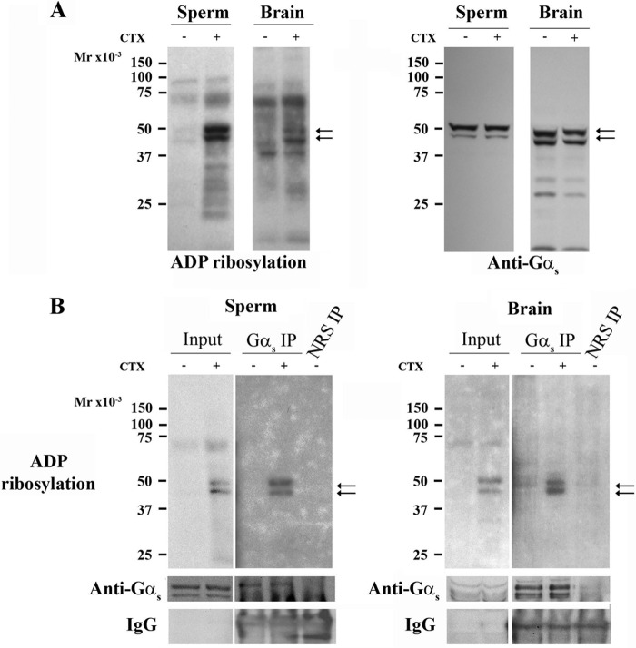
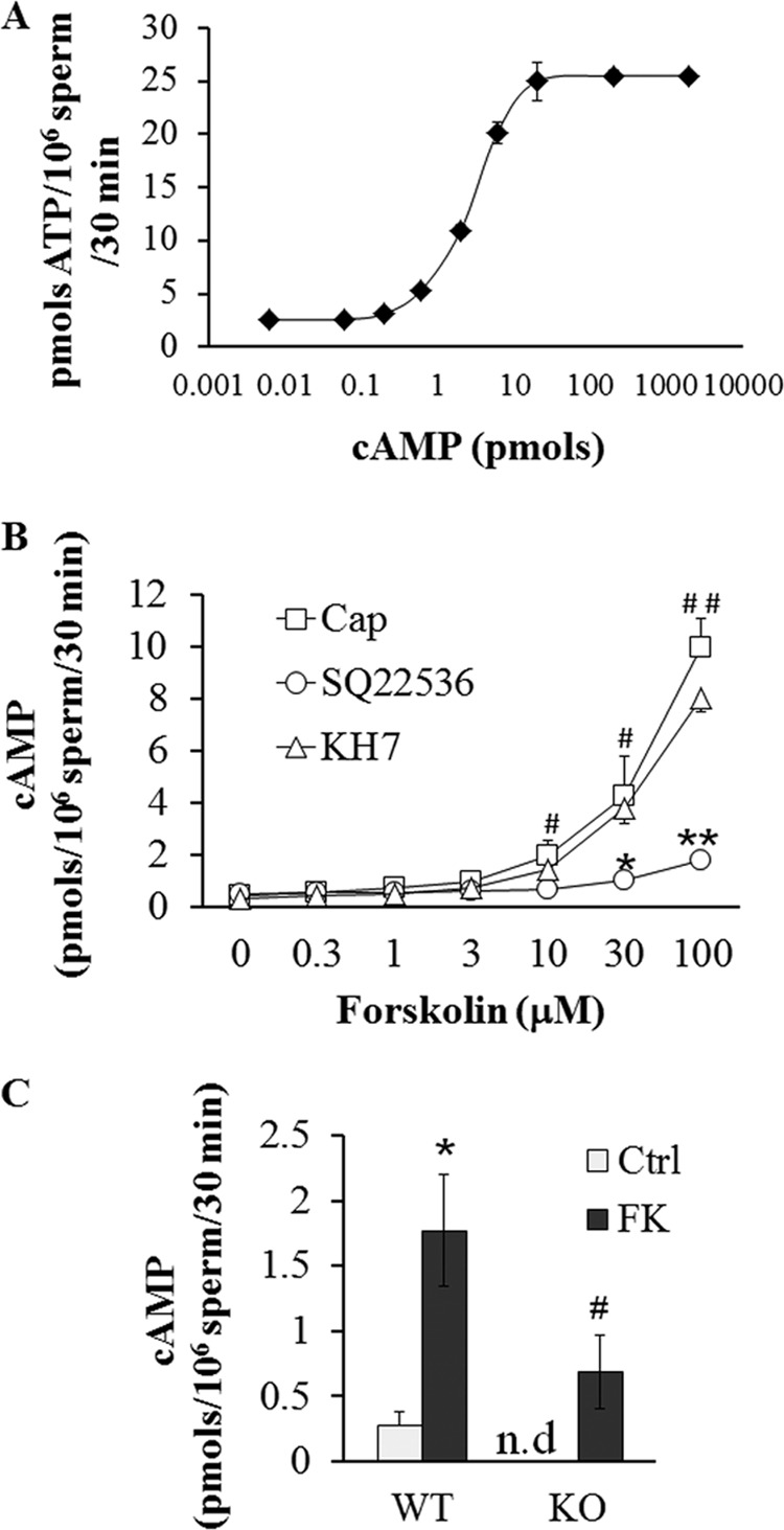
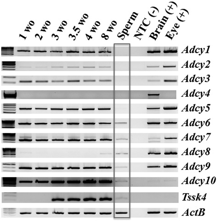
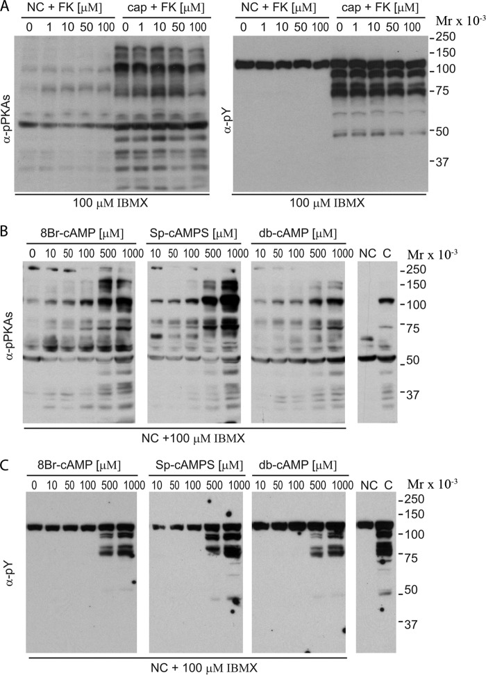
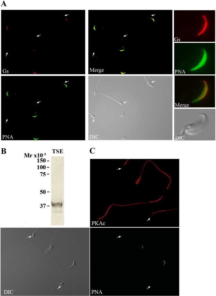
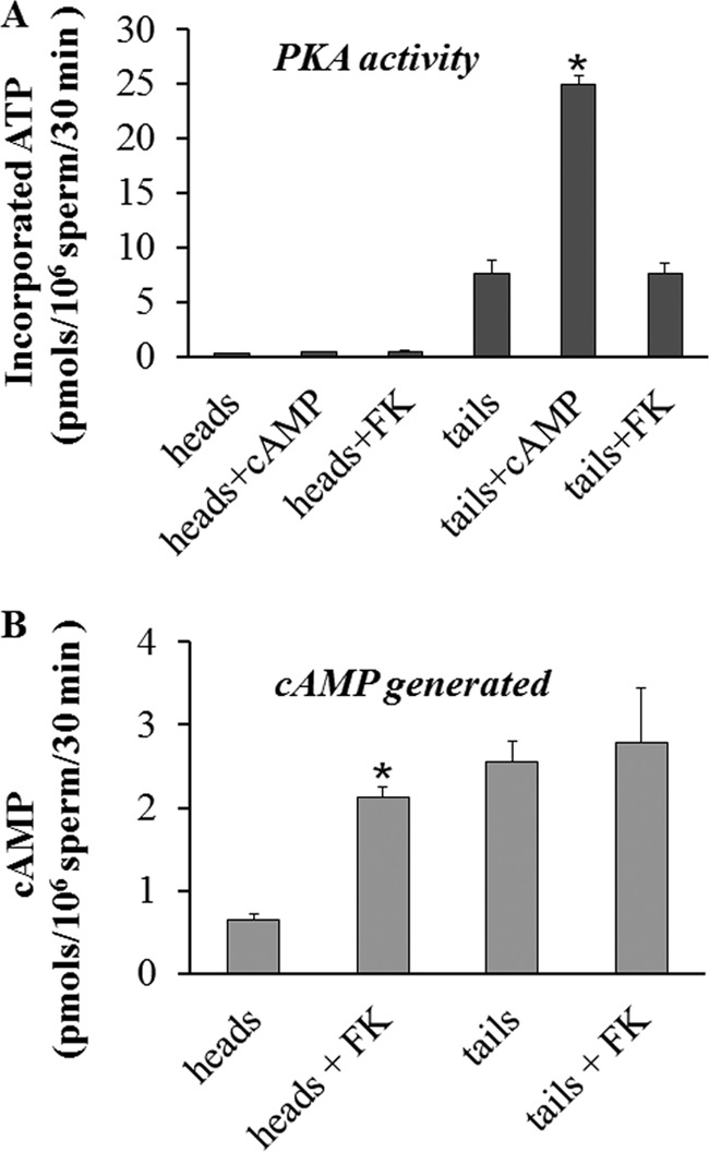
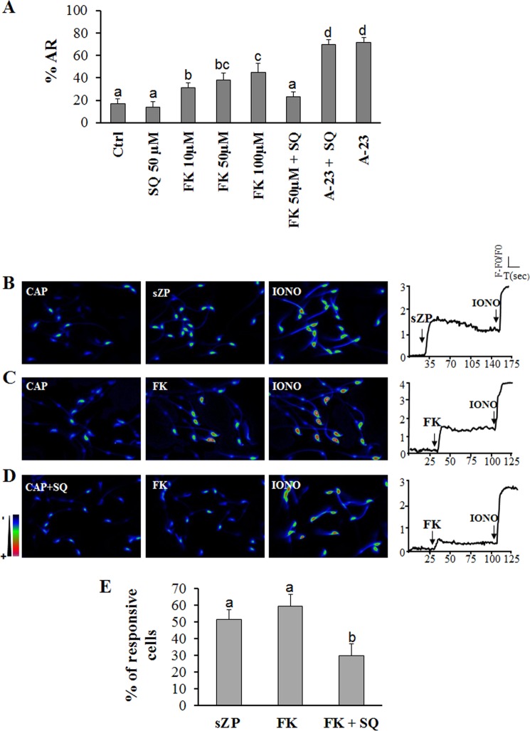
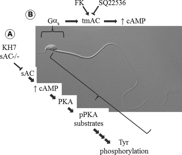
Fertilization competence is acquired in the female tract in a process known as capacitation. Capacitation is needed for the activation of motility (e.g. hyperactivation) and to prepare the sperm for an exocytotic process known as acrosome reaction. Although the HCO3(-)-dependent soluble adenylyl cyclase Adcy10 plays a role in motility, less is known about the source of cAMP in the sperm head. Transmembrane adenylyl cyclases (tmACs) are another possible source of cAMP. These enzymes are regulated by stimulatory heterotrimeric Gs proteins; however, the presence of Gs or tmACs in mammalian sperm has been controversial. In this study, we used Western blotting and cholera toxin-dependent ADP-ribosylation to show the Gs presence in the sperm head. Also, we showed that forskolin, a tmAC-specific activator, induces cAMP accumulation in sperm from both WT and Adcy10-null mice. This increase is blocked by the tmAC inhibitor SQ22536 but not by the Adcy10 inhibitor KH7. Although Gs immunoreactivity and tmAC activity are detected in the sperm head, PKA is only found in the tail, where Adcy10 was previously shown to reside. Consistent with an acrosomal localization, Gs reactivity is lost in acrosome-reacted sperm, and forskolin is able to increase intracellular Ca(2+) and induce the acrosome reaction. Altogether, these data suggest that cAMP pathways are compartmentalized in sperm, with Gs and tmAC in the head and Adcy10 and PKA in the flagellum.

Keywords: Acrosome Reaction; Adenylate Cyclase (Adenylyl Cyclase); Calcium Imaging; Cell Signaling; Cyclic AMP (cAMP); Forskolin; Heterotrimeric G Proteins; Protein Kinase A (PKA); Signal Transduction; Sperm Capacitation.


Figures

FIGURE 1.
FIGURE 1.
Stimulatory Gαs proteins are present in mouse sperm. A, mouse sperm and brain membranes were purified by differential centrifugation and assayed for in vitro [32P]ADP-ribosylation using pre-activated cholera toxin (CTX) as described under “Experimental Procedures.” After the reaction was completed, samples were subjected to SDS-PAGE, transferred to PVDF membranes, and exposed to autoradiography. Two bands were detected at 45 and 52 kDa that correspond to the molecular weight of Gαs (left panel). Western blots using a polyclonal anti-Gαs antibody were performed on the same membranes after autoradiography, and a doublet was detected at the same molecular weight as the autoradiography (right panel). B, immunoprecipitation using the same anti-Gαs antibody was carried out on both sperm and brain membrane extracts that had been previously subjected to cholera toxin [32P]ADP-ribosylation (Gαs immunoprecipitation). Immunoprecipitation controls were performed with normal rabbit serum (NRS IP). Immunoprecipitated samples were subjected to SDS-PAGE, transferred to PVDF membranes, exposed for autoradiography, and subsequently used for Western blot with anti-Gαs antibodies. IgG (light chain) in immunoprecipitation samples are also shown.
FIGURE 2.
FIGURE 2.
Forskolin increases cAMP levels in capacitated mouse sperm. A, representative cAMP concentration curve is shown, using known concentrations of cAMP. This curve was conducted in parallel for each independent assay. This method reproducibly detected cAMP concentrations ranging from 100 fmol to 20 pmol. In the absence of added cAMP, the basal value was 2.5 pmols of 32P incorporated into the Kemptide/30 min/106 sperm. B, sperm were incubated for 60 min in capacitating medium and further exposed to either 50 μm of KH7 or 100 μm SQ22536 for 15 min. Sperm samples were then treated with different concentrations of FK for 30 min as indicated. Each sample was processed for indirect quantification of cAMP using a PKA activity assay as described under “Experimental Procedures.” Two-way ANOVA followed by Bonferroni post tests were used to compare replicate means (n = 5). *, p < 0.05, and **, p < 0.01, when compared with capacitated sperm at same FK concentration. #, p < 0.05, and ##, p < 0.01, when compared with capacitated sperm with no FK. C, both WT and Adcy10 KO-capacitated sperm were exposed to 50 μm FK, and cAMP levels were analyzed using ELISA. Data represent mean ± S.E. n ≥4. Values for Adcy10 KO sperm before FK stimulation were below the detection limits and named in the graph as “nondetectable” (n.d.) (74). A paired t test was used to compare WT control versus WT + FK (*, p < 0.05), and a one-sample t test was used to determine whether the mean of KO + FK was different from the hypothetical value “0” (#, p < 0.05). Ctrl, control.
FIGURE 3.
FIGURE 3.
Different Adcy isoforms are expressed in mouse sperm and during testis development. Total RNA from sperm and testis of different developmental stages was isolated and used for RT-PCR experiments with isoform-specific intron-spanning primers. Eye and brain RNA were used as positive controls (+), and the absence of genomic contamination in the RNA samples was confirmed with reverse transcription negative controls (no reverse transcriptase) for each experiment (NTC(−)). Each isoform varies its pattern of expression during testis development. The expression of Adcy6–10 in sperm is observed.
FIGURE 4.
FIGURE 4.
FK has no effect on capacitation-associated phosphorylation events. A, sperm were incubated under noncapacitating (NC) and capacitating (cap) conditions in the presence of increasing concentrations of FK for 60 min. Protein extracts were analyzed by Western blotting with anti-Tyr(P) (αpY) and anti-phospho-PKA-substrate antibodies as described under “Experimental Procedures.” B and C, sperm were incubated in media without HCO3 supplemented with 100 μm IBMX and with increasing concentrations of 8-bromo-cAMP (8-Br-cAMP), (Sp)-cAMPS, or dibutyryl cyclic AMP (db-cAMP). Samples were processed for Western blotting with anti-pPKAs (B), stripped, and further processed with anti-Tyr(P)antibodies (C). All Western blots are representative of experiments repeated at least three times.
FIGURE 5.
FIGURE 5.
s and the PKA catalytic subunit localized to different sperm compartments. A, for immunofluorescence assays, mouse sperm were air-dried, fixed, permeabilized, and probed with a polyclonal anti-Gαs antibody. PNA was used to follow the acrosomal status. Anti-Gαs (red) and PNA (green) labeling showed that Gαs is lost during acrosomal reaction (merged color panels). Differential interference contrast (DIC) microscopy was used to control for sperm morphology. Images are representative of at least three experimental replicates. B, immunodetection of the catalytic subunit of PKA by Western blotting. Total sperm extracts (TSE) were subjected to SDS-PAGE, transferred to PVDF membranes, and analyzed by Western blotting with anti-PKA catalytic (PKAc) subunit antibodies. Only one band of the expected molecular weight is observed. C, immunolocalization of cPKA in mature sperm. Immunofluorescence was carried out in fixed and permeabilized sperm as detailed under “Experimental Procedures.” Anti-cPKA staining of whole sperm shows the presence of PKA only in the sperm flagellum.
FIGURE 6.
FIGURE 6.
PKA activity is only observed in sperm tails, whereas FK-induced cAMP production is detected in sperm heads. A, head and tail fractions from capacitated sperm were separated as described under “Experimental Procedures” and assayed for PKA activity using [32P]ATP and Kemptide as substrate in the presence of either 1 mm cAMP or 100 μm FK. Data represent mean ± S.E. of five independent experiments performed in triplicates; two-way ANOVA followed by Bonferroni post tests were used to compare replicate means. *, p < 0.001 when compared with tails without cAMP addition. B, head and tails fractions from capacitated sperm were treated with 100 μm FK and processed for cAMP quantification using PKA activity assays as described under “Experimental Procedures.” Data represent mean ± S.E. of five independent experiments performed in triplicate; two-way ANOVA followed by Bonferroni post tests were used to compare replicate means. *, p < 0.05 when compared with heads without FK addition.
FIGURE 7.
FIGURE 7.
FK promotes acrosome reaction, and [Ca2+]i increases in capacitated mouse sperm. A, FK induces acrosome reaction. Mouse sperm were incubated under capacitating conditions for 1 h and then further incubated for 30 min in the absence or presence of the tmAC inhibitor SQ22536. Subsequently, either FK or calcium ionophore A23187 (A-23) was added, and sperm were incubated for an additional 30 min. Acrosomal status was assessed as explained under “Experimental Procedures.” Percentage of acrosome-reacted sperm (%AR) was calculated based on seven independent experiments run in triplicate. B, pseudocolored fluorescence images illustrating [Ca2+]i levels before (CAP) and after addition of sZP to capacitated mouse sperm. The [Ca2+]i increase induced subsequently by 10 μm ionomycin (IONO) is also shown as a positive control. The right panel shows representative [Ca2+]i traces of sperm subjected to the conditions described above. Arrows indicate agonist application. C, fluorescence images of [Ca2+]i levels before (CAP) and after addition of 50 μm FK (FK) to capacitated mouse sperm. D, fluorescence images of [Ca2+]i of capacitated mouse sperm incubated for 30 min with the tmAC inhibitor SQ22536 (CAP+SQ) and after the addition of FK. E, percentage of sperm displaying [Ca2+]i increases in response to ZP compared with those responsive to FK alone or after incubation with SQ22536. The number of cells analyzed for each condition is ZP = 121, FK = 197, FK + SQ = 176 (each experimental condition was performed in at least four independent experiments). Tukey's multiple comparison statistics tests were performed for both acrosome reaction and [Ca2+]i experiments. Means of groups that have different letters differ significantly (p < 0.01).
FIGURE 8.
FIGURE 8.
Proposed model for the spatial distribution of cAMP in sperm functions. A, Adcy10 mediates activation of flagellum cAMP-dependent pathways such as the activation of PKA is upstream of the increase in tyrosine phosphorylation. B, Gαs is in the head and activates a tmAC that increases cAMP levels that would be necessary to activate acrosome reaction by FK. Inhibition of tmAC by SQ22536 blocks the FK-induced acrosome reaction. The cAMP effector in the head is not PKA. One possible cAMP target in the sperm head is the G protein exchange factor called EPAC (48).

Similar articles

Cited by

References

    1. Visconti P. E., Krapf D., de la Vega-Beltrán JL, Acevedo J. J., Darszon A. (2011) Ion channels, phosphorylation and mammalian sperm capacitation. Asian J. Androl. 13, 395–405 - PMC - PubMed
    1. Bailey J. L. (2010) Factors regulating sperm capacitation. Syst. Biol. Reprod. Med. 56, 334–348 - PubMed
    1. Visconti P. E., Galantino-Homer H., Moore G. D., Bailey J. L., Ning X., Fornes M., Kopf G. S. (1998) The molecular basis of sperm capacitation. J. Androl. 19, 242–248 - PubMed
    1. Morgan D. J., Weisenhaus M., Shum S., Su T., Zheng R., Zhang C., Shokat K. M., Hille B., Babcock D. F., McKnight G. S. (2008) Tissue-specific PKA inhibition using a chemical genetic approach and its application to studies on sperm capacitation. Proc. Natl. Acad. Sci. U.S.A. 105, 20740–20745 - PMC - PubMed
    1. Nolan M. A., Babcock D. F., Wennemuth G., Brown W., Burton K. A., McKnight G. S. (2004) Sperm-specific protein kinase A catalytic subunit Cα2 orchestrates cAMP signaling for male fertility. Proc. Natl. Acad. Sci. U.S.A. 101, 13483–13488 - PMC - PubMed

Publication types

MeSH terms

LinkOut - more resources