

Two actin-interacting protein 1 isoforms function redundantly in the somatic gonad and are essential for reproduction in Caenorhabditis elegans
- PMID: 24130131
- PMCID: PMC4082699
- DOI: 10.1002/cm.21152
Two actin-interacting protein 1 isoforms function redundantly in the somatic gonad and are essential for reproduction in Caenorhabditis elegans
Abstract
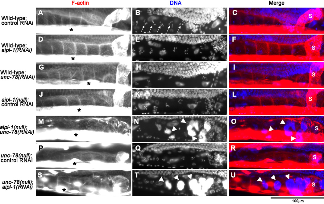
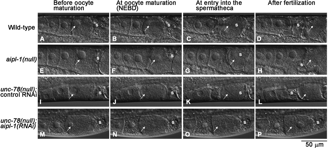
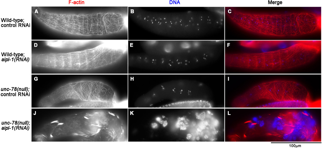
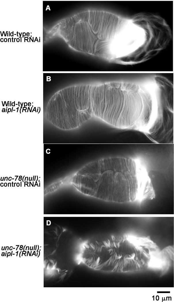
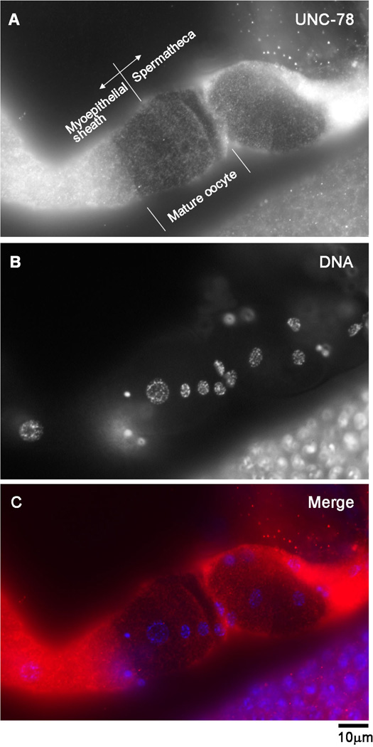
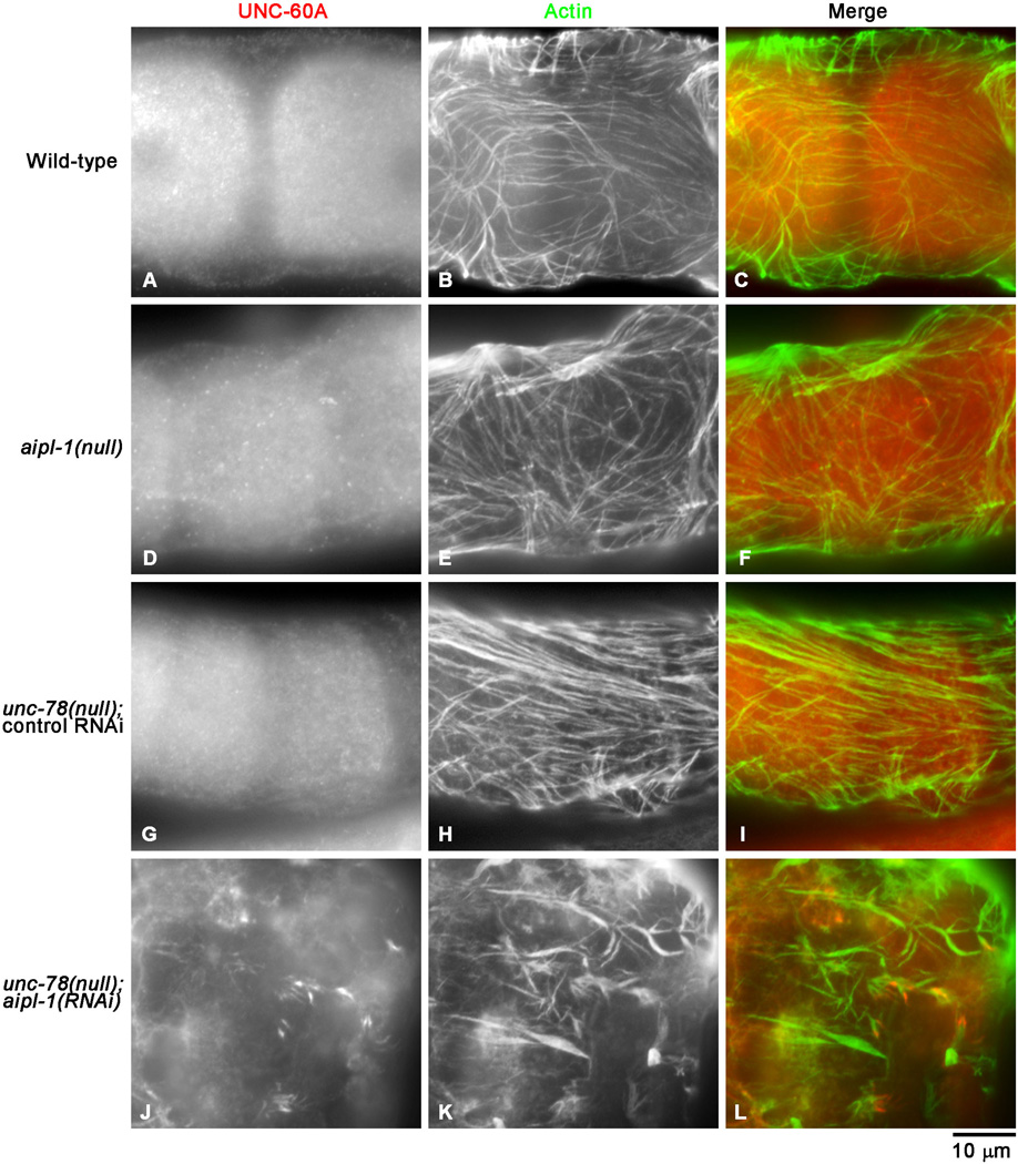
The somatic gonad of the nematode Caenorhabditis elegans exhibits highly regulated contractility during ovulation, which is essential for successful reproduction. Nonstriated actin filament networks in the myoepithelial sheath at the proximal ovary provide contractile forces to push a mature oocyte for ovulation, but the mechanism of assembly and regulation of the contractile actin networks is poorly understood. Here, we show that actin-interacting protein 1 (AIP1) is essential for the assembly of the contractile actin networks in the myoepithelial sheath. AIP1 promotes disassembly of actin filaments in the presence of actin depolymerizing factor (ADF)/cofilin. C. elegans has two AIP1 genes, unc-78 and aipl-1. Mutation or RNA interference of a single AIP1 isoform causes only minor impacts on reproduction. However, simultaneous depletion of the two AIP1 isoforms causes sterility. AIP1-depleted animals show very weak contractility of the myoepithelial sheath and fail to ovulate a mature oocyte, which results in accumulation of endomitotic oocytes in the ovary. Depletion of AIP1 prevents assembly of actin networks and causes abnormal aggregation of actin as well as ADF/cofilin in the myoepithelial sheath. These results indicate that two AIP1 isoforms have redundant roles in assembly of the contractile apparatuses necessary for C. elegans reproduction.
Keywords: actin depolymerizing factor/cofilin; actin dynamics; myoepithelial cells; ovulation; spermatheca; sterility.
Copyright © 2013 Wiley Periodicals, Inc.
Figures
Similar articles
-
Essential role of ADF/cofilin for assembly of contractile actin networks in the C. elegans somatic gonad.J Cell Sci. 2008 Aug 15;121(Pt 16):2662-70. doi: 10.1242/jcs.034215. Epub 2008 Jul 24. J Cell Sci. 2008. PMID: 18653537 Free PMC article.
-
The two actin-interacting protein 1 genes have overlapping and essential function for embryonic development in Caenorhabditis elegans.Mol Biol Cell. 2011 Jul 1;22(13):2258-69. doi: 10.1091/mbc.E10-12-0934. Epub 2011 May 5. Mol Biol Cell. 2011. PMID: 21551072 Free PMC article.
-
Actin-interacting Protein 1 Promotes Disassembly of Actin-depolymerizing Factor/Cofilin-bound Actin Filaments in a pH-dependent Manner.J Biol Chem. 2016 Mar 4;291(10):5146-56. doi: 10.1074/jbc.M115.713495. Epub 2016 Jan 8. J Biol Chem. 2016. PMID: 26747606 Free PMC article.
-
Functions of actin-interacting protein 1 (AIP1)/WD repeat protein 1 (WDR1) in actin filament dynamics and cytoskeletal regulation.Biochem Biophys Res Commun. 2018 Nov 25;506(2):315-322. doi: 10.1016/j.bbrc.2017.10.096. Epub 2017 Oct 19. Biochem Biophys Res Commun. 2018. PMID: 29056508 Free PMC article. Review.
-
Regulation of actin filament dynamics by actin depolymerizing factor/cofilin and actin-interacting protein 1: new blades for twisted filaments.Biochemistry. 2003 Nov 25;42(46):13363-70. doi: 10.1021/bi034600x. Biochemistry. 2003. PMID: 14621980 Review.
Cited by
-
Wdr1-mediated cell shape dynamics and cortical tension are essential for epidermal planar cell polarity.Nat Cell Biol. 2015 May;17(5):592-604. doi: 10.1038/ncb3146. Epub 2015 Apr 27. Nat Cell Biol. 2015. PMID: 25915128 Free PMC article.
-
INF2- and FHOD-related formins promote ovulation in the somatic gonad of C. elegans.Cytoskeleton (Hoboken). 2016 Dec;73(12):712-728. doi: 10.1002/cm.21341. Epub 2016 Nov 9. Cytoskeleton (Hoboken). 2016. PMID: 27770600 Free PMC article.
-
Aip1 promotes actin filament severing by cofilin and regulates constriction of the cytokinetic contractile ring.J Biol Chem. 2015 Jan 23;290(4):2289-300. doi: 10.1074/jbc.M114.612978. Epub 2014 Dec 1. J Biol Chem. 2015. PMID: 25451933 Free PMC article.
-
Regulation of Actin Dynamics in the C. elegans Somatic Gonad.J Dev Biol. 2019 Mar 20;7(1):6. doi: 10.3390/jdb7010006. J Dev Biol. 2019. PMID: 30897735 Free PMC article. Review.
-
Spectrin regulates cell contractility through production and maintenance of actin bundles in the Caenorhabditis elegans spermatheca.Mol Biol Cell. 2018 Oct 1;29(20):2433-2449. doi: 10.1091/mbc.E18-06-0347. Epub 2018 Aug 9. Mol Biol Cell. 2018. PMID: 30091661 Free PMC article.
References
-
- Aizawa H, Katadae M, Maruya M, Sameshima M, Murakami-Murofushi K, Yahara I. Hyperosmotic stress-induced reorganization of actin bundles in Dictyostelium cells over-expressing cofilin. Genes Cells. 1999;4:311–324. - PubMed
Publication types
MeSH terms
Substances
Grants and funding
LinkOut - more resources
Full Text Sources
Other Literature Sources
Research Materials
Miscellaneous
