

Vasculogenesis in experimental stroke after human cerebral endothelial cell transplantation
- PMID: 24130140
- PMCID: PMC4083631
- DOI: 10.1161/STROKEAHA.113.001943
Vasculogenesis in experimental stroke after human cerebral endothelial cell transplantation
Abstract
Background and purpose: Despite the reported functional recovery in transplanted stroke models and patients, the mechanism of action underlying stem cell therapy remains not well understood. Here, we examined the role of stem cell-mediated vascular repair in stroke.
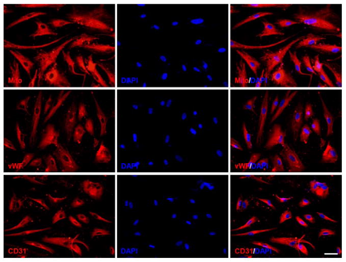
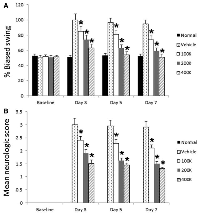
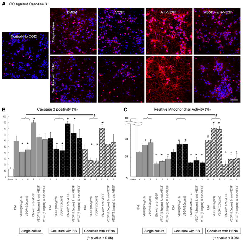
Methods: Adult rats were exposed to transient occlusion of the middle cerebral artery and 3 hours later randomly stereotaxically transplantated with 100K, 200K, or 400K human cerebral endothelial cell 6 viable cells or vehicle. Animals underwent neurological examination and motor test up to day 7 after transplantation then euthanized for immunostaining against neuronal, vascular, and specific human antigens. A parallel in vitro study cocultured rat primary neuronal cells with human cerebral endothelial cell 6 under oxygen-glucose deprivation and treated with vascular endothelial growth factor (VEGF) and anti-VEGF.
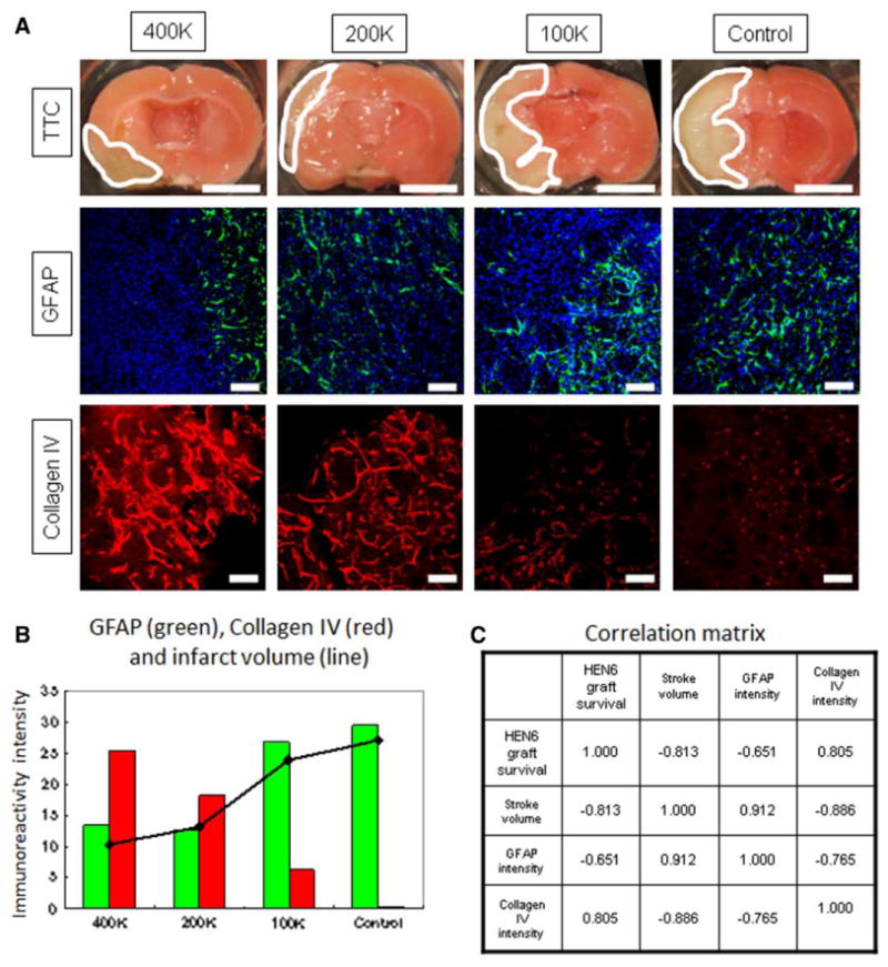
Results: Stroke animals that received vehicle infusion displayed typical occlusion of the middle cerebral artery-induced behavioral impairments that were dose-dependently reduced in transplanted stroke animals at days 3 and 7 after transplantation and accompanied by increased expression of host neuronal and vascular markers adjacent to the transplanted cells. Some transplanted cells showed a microvascular phenotype and juxtaposed to the host vasculature. Infarct volume in transplanted stroke animals was significantly smaller than vehicle-infused stroke animals. Moreover, rat neurons cocultured with human cerebral endothelial cell 6 or treated with VEGF exhibited significantly less oxygen-glucose deprivation-induced cell death that was blocked by anti-VEGF treatment.
Conclusions: We found attenuation of behavioral and histological deficits coupled with robust vasculogenesis and neurogenesis in endothelial cell-transplanted stroke animals, suggesting that targeting vascular repair sets in motion a regenerative process in experimental stroke possibly via the VEGF pathway.
Keywords: endothelial cells; neurogenesis; stem cells; stroke.
Figures

Similar articles
-
The neural stem cell line CTX0E03 promotes behavioral recovery and endogenous neurogenesis after experimental stroke in a dose-dependent fashion.Neurorehabil Neural Repair. 2009 Nov;23(9):895-909. doi: 10.1177/1545968309335978. Epub 2009 Jul 24. Neurorehabil Neural Repair. 2009. PMID: 19633272
-
Transplantation of Flk-1+ human bone marrow-derived mesenchymal stem cells promotes angiogenesis and neurogenesis after cerebral ischemia in rats.Eur J Neurosci. 2011 Jul;34(1):87-98. doi: 10.1111/j.1460-9568.2011.07733.x. Epub 2011 Jun 21. Eur J Neurosci. 2011. PMID: 21692879
-
Allogeneic adipose-derived mesenchymal stem cell sheet that produces neurological improvement with angiogenesis and neurogenesis in a rat stroke model.J Neurosurg. 2019 Feb 22;132(2):442-455. doi: 10.3171/2018.11.JNS182331. Print 2020 Feb 1. J Neurosurg. 2019. PMID: 30797215
-
Nose-to-brain delivery of stem cells in stroke: the role of extracellular vesicles.Stem Cells Transl Med. 2024 Nov 12;13(11):1043-1052. doi: 10.1093/stcltm/szae072. Stem Cells Transl Med. 2024. PMID: 39401332 Free PMC article. Review.
-
Stem cell therapy in stroke.Cell Mol Life Sci. 2009 Mar;66(5):757-72. doi: 10.1007/s00018-008-8346-1. Cell Mol Life Sci. 2009. PMID: 18989624 Free PMC article. Review.
Cited by
-
Probing Multiple Transplant Delivery Routes of CD+34 Stem Cells for Promoting Behavioral and Histological Benefits in Experimental Ischemic Stroke.Stem Cells Transl Med. 2024 Feb 14;13(2):177-190. doi: 10.1093/stcltm/szad081. Stem Cells Transl Med. 2024. PMID: 38016184 Free PMC article.
-
Improvement of erectile dysfunction using endothelial progenitor cells from fetal cerebral vasculature in the cavernous nerve injury of rats.Basic Clin Androl. 2022 Dec 1;32(1):21. doi: 10.1186/s12610-022-00171-x. Basic Clin Androl. 2022. PMID: 36451096 Free PMC article.
-
Stem Cell Recipes of Bone Marrow and Fish: Just What the Stroke Doctors Ordered.Stem Cell Rev Rep. 2017 Apr;13(2):192-197. doi: 10.1007/s12015-016-9716-y. Stem Cell Rev Rep. 2017. PMID: 28064388 Review.
-
HIF‑1α in cerebral ischemia (Review).Mol Med Rep. 2022 Feb;25(2):41. doi: 10.3892/mmr.2021.12557. Epub 2021 Dec 8. Mol Med Rep. 2022. PMID: 34878158 Free PMC article. Review.
-
Proteomic Analysis of a Rat Cerebral Ischemic Injury Model after Human Cerebral Endothelial Cell Transplantation.J Korean Neurosurg Soc. 2016 Nov;59(6):544-550. doi: 10.3340/jkns.2016.59.6.544. Epub 2016 Oct 24. J Korean Neurosurg Soc. 2016. PMID: 27847565 Free PMC article.
References
-
- Stem Cell Therapies as an Emerging Paradigm in Stroke Participants. Stem cell therapies as an emerging paradigm in stroke (steps): bridging basic and clinical science for cellular and neurogenic factor therapy in treating stroke. Stroke. 2009;40:510–515. - PubMed
-
- Kondziolka D, Wechsler L, Goldstein S, Meltzer C, Thulborn KR, Gebel J, et al. Transplantation of cultured human neuronal cells for patients with stroke. Neurology. 2000;55:565–569. - PubMed
-
- Jeong SW, Chu K, Jung KH, Kim SU, Kim M, Roh JK. Human neural stem cell transplantation promotes functional recovery in rats with experimental intracerebral hemorrhage. Stroke. 2003;34:2258–2263. - PubMed
Publication types
MeSH terms
Grants and funding
LinkOut - more resources
Full Text Sources
Other Literature Sources
Medical
