. 2013 Oct;25(10):4101-9.
doi: 10.1105/tpc.113.117184. Epub 2013 Oct 15.

Plant purine nucleoside catabolism employs a guanosine deaminase required for the generation of xanthosine in Arabidopsis

Affiliations

Plant purine nucleoside catabolism employs a guanosine deaminase required for the generation of xanthosine in Arabidopsis

Kathleen Dahncke et al. Plant Cell. 2013 Oct.

Abstract

Purine nucleotide catabolism is common to most organisms and involves a guanine deaminase to convert guanine to xanthine in animals, invertebrates, and microorganisms. Using metabolomic analysis of mutants, we demonstrate that Arabidopsis thaliana uses an alternative catabolic route employing a highly specific guanosine deaminase (GSDA) not reported from any organism so far. The enzyme is ubiquitously expressed and deaminates exclusively guanosine and 2'-deoxyguanosine but no other aminated purines, pyrimidines, or pterines. GSDA belongs to the cytidine/deoxycytidylate deaminase family of proteins together with a deaminase involved in riboflavin biosynthesis, the chloroplastic tRNA adenosine deaminase Arg and a predicted tRNA-specific adenosine deaminase 2 in A. thaliana. GSDA is conserved in plants, including the moss Physcomitrella patens, but is absent in the algae and outside the plant kingdom. Our data show that xanthosine is exclusively generated through the deamination of guanosine by GSDA in A. thaliana, excluding other possible sources like the dephosphorylation of xanthosine monophosphate. Like the nucleoside hydrolases NUCLEOSIDE HYDROLASE1 (NSH1) and NSH2, GSDA is located in the cytosol, indicating that GMP catabolism to xanthine proceeds in a mostly cytosolic pathway via guanosine and xanthosine. Possible implications for the biosynthetic route of purine alkaloids (caffeine and theobromine) and ureides in other plants are discussed.


Figures
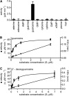
Figure 1.
Figure 1.
Enzymatic Activity and Determination of Kinetic Constants for GSDA. (A) Enzymatic activity of GSDA with 1 mM different nucleotides, nucleosides, nucleobases, and pterines as substrates. THF, tetrahydrofolate. Error bars are sd (n = 3). (B) Left axis, determination of catalytic velocity (v) in terms of ammonia production at different guanosine concentrations fitted using the Michaelis-Menten equation (R2 = 0.9343). Right axis, Hanes plot: ratio of guanosine concentration and velocity (S/v) plotted against guanosine concentration (S) fitted by linear regression (r2 = 0.9936). Error bars are sd (n = 5). (C) As in (B) but using 2’-deoxyguanosine as substrate (R2 = 0.9043, r2 = 0.9698, n = 4).
Figure 2.
Figure 2.
Characterization of Mutants. (A) The genomic organization of the GSDA gene. Boxes represent the exonic regions of the coding sequence. The position of the T-DNA insertions in the respective mutant plant lines are indicated by triangles. Numbers indicate the distance from the first base altered by the insertion to the last base of the stop codon. (B) Relative quantification of GSDA transcript in seedlings of the wild type and the two mutant lines by quantitative PCR using UBIQUITIN10 as the reference gene. Values have been normalized to mean value of corresponding wild-type samples. Error bars are sd (n = 3; three independent RNA extracts were prepared from a pool of seedlings for each line and used for cDNA synthesis and quantitative PCR). Col-0, Columbia-0; n.d., not detectable. (C) Assessment of GSDA protein in seedlings of the wild type and the two mutant lines as well as different organs of the wild type by immunoblot employing GSDA-specific antiserum. (D) Analysis of metabolite extracts using HPLC with photometric detection from dry seeds of the wild type, the two gsda mutants, two complementation lines containing either a Strep-tagged or YFP-tagged transgene, the nsh1-1 mutant, and a double mutant of gsda-2 and nsh1-1. mAU, milliabsorption units.
Figure 3.
Figure 3.
Metabolite Quantification in Different Tissues of Single and Double Mutants. (A) Concentration of guanosine, xanthosine, and uridine in gsda-2, nsh1-1, and the gsda-2 nsh1-1 double mutant in rosette leaves, roots, and yellow siliques of 10-week-old plants grown under long-day conditions. Error bars are sd (n = 5). fw, fresh weight. (B) Assessment of GSDA protein in corresponding wild-type samples by immunoblot employing GSDA-specific antiserum (8 μg total protein per lane). Col-0, Columbia-0.
Figure 4.
Figure 4.
Subcellular Localization of GSDA. (A) to (C) Confocal fluorescence microscopy images of lower leaf epidermis cells of N. benthamiana transiently expressing N-terminally YFP-tagged GSDA (YFP-GSDA) and C-terminally CFP-tagged cytosolic protein (β-UP-CFP). YFP (A), CFP (B), and YFP + CFP (C) fluorescence, respectively. Bar = 20 μm. (D) and (E) Mesophyll cell protoplast of lines carrying a YFP-GSDA transgene. YFP fluorescence (D) and YFP + chlorophyll autofluorescence (E). Bar = 10 μm. (F) Stability of the YFP-GSDA fusion protein after transient expression in N. benthamiana analyzed by immunoblot developed with a GFP-specific antibody.
Figure 5.
Figure 5.
Phylogenetic Analysis of the GDA/GSDA Protein Subfamily. The sequence alignment of Supplemental Figure 8 online (available as Supplemental Data Set 1 online) was used to construct a phylogenetic tree employing the MEGA5 software (maximum likelihood, WAG+G model, nearest neighbor interchange). A consensus tree from 1000 bootstraps was constructed. Only bootstrap values above 70 are shown. The GSDA portion of the tree was amplified 5 times to better visualize the branching pattern. Eukaryotes within the GDA portion of the tree are indicated by dark background.
Figure 6.
Figure 6.
Model of the Metabolic Pathway for GMP Degradation in A. thaliana. GMP is dephosphorylated by a so far unknown phosphatase (PPase) to guanosine, which is either deaminated to xanthosine by GSDA or salvaged into nucleotides and nucleic acids. The hydrolysis of xanthosine to xanthine and Rib is catalyzed by NSH1 possibly with the participation of NSH2. In general, xanthosine and xanthine cannot be salvaged and are destined for degradation via purine ring catabolism (gray shaded area). Neither XMP nor inosine gives rise to xanthosine in vivo.

Similar articles

Cited by

References

    1. Alonso J.M., et al. (2003). Genome-wide insertional mutagenesis of Arabidopsis thaliana. Science 301: 653–657 - PubMed
    1. Ashihara H. (2012). Xanthosine metabolism in plants: Metabolic fate of exogenously supplied C-14-labelled xanthosine and xanthine in intact mungbean seedlings. Phytochem. Lett. 5: 100–103
    1. Ashihara H., Sano H., Crozier A. (2008). Caffeine and related purine alkaloids: Biosynthesis, catabolism, function and genetic engineering. Phytochemistry 69: 841–856 - PubMed
    1. Ashihara H., Takasawa Y., Suzuki T. (1997). Metabolic fate of guanosine in higher plants. Physiol. Plant. 100: 909–916
    1. Brychkova G., Alikulov Z., Fluhr R., Sagi M. (2008). A critical role for ureides in dark and senescence-induced purine remobilization is unmasked in the Atxdh1 Arabidopsis mutant. Plant J. 54: 496–509 - PubMed

Publication types

Associated data