Metformin targets c-MYC oncogene to prevent prostate cancer
- PMID: 24130167
- PMCID: PMC3845895
- DOI: 10.1093/carcin/bgt307
Metformin targets c-MYC oncogene to prevent prostate cancer
Abstract
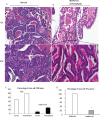
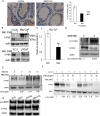
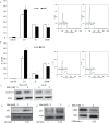
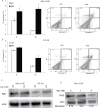
Prostate cancer (PCa) is the second leading cause of cancer-related death in American men and many PCa patients develop skeletal metastasis. Current treatment modalities for metastatic PCa are mostly palliative with poor prognosis. Epidemiological studies indicated that patients receiving the diabetic drug metformin have lower PCa risk and better prognosis, suggesting that metformin may have antineoplastic effects. The mechanism by which metformin acts as chemopreventive agent to impede PCa initiation and progression is unknown. The amplification of c-MYC oncogene plays a key role in early prostate epithelia cell transformation and PCa growth. The purpose of this study is to investigate the effect of metformin on c-myc expression and PCa progression. Our results demonstrated that (i) in Hi-Myc mice that display murine prostate neoplasia and highly resemble the progression of human prostate tumors, metformin attenuated the development of prostate intraepithelial neoplasia (PIN, the precancerous lesion of prostate) and PCa lesions. (ii) Metformin reduced c-myc protein levels in vivo and in vitro. In Myc-CaP mouse PCa cells, metformin decreased c-myc protein levels by at least 50%. (iii) Metformin selectively inhibited the growth of PCa cells by stimulating cell cycle arrest and apoptosis without affecting the growth of normal prostatic epithelial cells (RWPE-1). (iv) Reduced PIN formation by metformin was associated with reduced levels of androgen receptor and proliferation marker Ki-67 in Hi-Myc mouse prostate glands. Our novel findings suggest that by downregulating c-myc, metformin can act as a chemopreventive agent to restrict prostatic neoplasia initiation and transformation.
Summary: Metformin, an old antidiabetes drug, may inhibit prostate intraepithelial neoplasia transforming to cancer lesion via reducing c-MYC, an 'old' overexpressed oncogene. This study explores chemopreventive efficacy of metformin in prostate cancer and its link to cMYC in vitro and in vivo.
Figures


Similar articles
-
MYC overexpression induces prostatic intraepithelial neoplasia and loss of Nkx3.1 in mouse luminal epithelial cells.PLoS One. 2010 Feb 25;5(2):e9427. doi: 10.1371/journal.pone.0009427. PLoS One. 2010. PMID: 20195545 Free PMC article.
-
Preneoplastic prostate lesions: an opportunity for prostate cancer prevention.Ann N Y Acad Sci. 2001 Dec;952:135-44. doi: 10.1111/j.1749-6632.2001.tb02734.x. Ann N Y Acad Sci. 2001. PMID: 11795433 Review.
-
Caveolin-1 upregulation contributes to c-Myc-induced high-grade prostatic intraepithelial neoplasia and prostate cancer.Mol Cancer Res. 2012 Feb;10(2):218-29. doi: 10.1158/1541-7786.MCR-11-0451. Epub 2011 Dec 5. Mol Cancer Res. 2012. PMID: 22144662 Free PMC article.
-
SPOP regulates prostate epithelial cell proliferation and promotes ubiquitination and turnover of c-MYC oncoprotein.Oncogene. 2017 Aug 17;36(33):4767-4777. doi: 10.1038/onc.2017.80. Epub 2017 Apr 17. Oncogene. 2017. PMID: 28414305 Free PMC article.
-
Apoptotic regulators in prostatic intraepithelial neoplasia (PIN): value in prostate cancer detection and prevention.Prostate Cancer Prostatic Dis. 2005;8(1):7-13. doi: 10.1038/sj.pcan.4500757. Prostate Cancer Prostatic Dis. 2005. PMID: 15477876 Review.
Cited by
-
Metformin use and hospital attendance-related resources utilization among diabetic patients with prostate cancer on androgen deprivation therapy: A population-based cohort study.Cancer Med. 2023 Apr;12(8):9128-9132. doi: 10.1002/cam4.5651. Epub 2023 Feb 3. Cancer Med. 2023. PMID: 36734312 Free PMC article.
-
Metformin alters H2A.Z dynamics and regulates androgen dependent prostate cancer progression.Oncotarget. 2018 Dec 11;9(97):37054-37068. doi: 10.18632/oncotarget.26457. eCollection 2018 Dec 11. Oncotarget. 2018. PMID: 30651935 Free PMC article.
-
Synergism between metformin and statins in modifying the risk of biochemical recurrence following radical prostatectomy in men with diabetes.Prostate Cancer Prostatic Dis. 2015 Mar;18(1):63-8. doi: 10.1038/pcan.2014.47. Epub 2014 Nov 18. Prostate Cancer Prostatic Dis. 2015. PMID: 25403419
-
Metformin Decreases 2-HG Production through the MYC-PHGDH Pathway in Suppressing Breast Cancer Cell Proliferation.Metabolites. 2021 Jul 26;11(8):480. doi: 10.3390/metabo11080480. Metabolites. 2021. PMID: 34436421 Free PMC article.
-
GLS-driven glutamine catabolism contributes to prostate cancer radiosensitivity by regulating the redox state, stemness and ATG5-mediated autophagy.Theranostics. 2021 Jun 26;11(16):7844-7868. doi: 10.7150/thno.58655. eCollection 2021. Theranostics. 2021. PMID: 34335968 Free PMC article.
References
-
- Jemal A., et al. (2010). Cancer statistics, 2010. CA Cancer J. Clin., 60, 277–300 - PubMed
-
- SEER (2012). SEER Stat Fact Sheets: Prostate http://seer.cancer.gov/statfacts/html/prost.html (31 January 2013, date last accessed).
-
- Moser L., et al. (2008). Hormone-refractory and metastatic prostate cancer—palliative radiotherapy. Front. Radiat. Ther. Oncol., 41, 117–125 - PubMed
-
- Bostwick D.G. (1988). Premalignant lesions of the prostate. Semin. Diagn. Pathol., 5, 240–253 - PubMed
-
- Colanzi P., et al. (1998). Prostatic intraepithelial neoplasia and prostate cancer: analytical evaluation. Adv. Clin. Path., 2, 271–284 - PubMed
Publication types
MeSH terms
Substances
Grants and funding
LinkOut - more resources
Full Text Sources
Other Literature Sources
Medical
Molecular Biology Databases
Miscellaneous

