Review
. 2013 Sep;36(3):308-15.
doi: 10.1590/S1415-47572013000300002. Epub 2013 Aug 30.

Replicating animal mitochondrial DNA

Affiliations
Review

Replicating animal mitochondrial DNA

Emily A McKinney et al. Genet Mol Biol. 2013 Sep.

Abstract

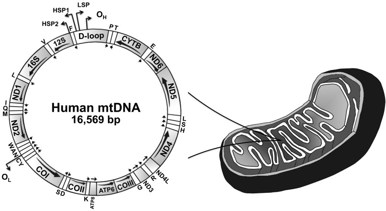
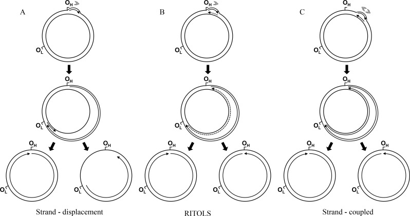
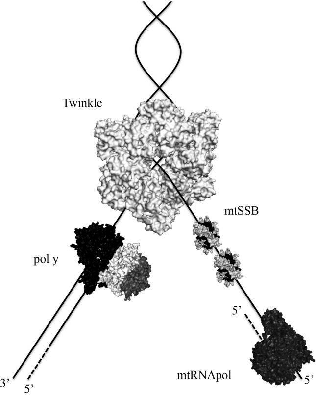
The field of mitochondrial DNA (mtDNA) replication has been experiencing incredible progress in recent years, and yet little is certain about the mechanism(s) used by animal cells to replicate this plasmid-like genome. The long-standing strand-displacement model of mammalian mtDNA replication (for which single-stranded DNA intermediates are a hallmark) has been intensively challenged by a new set of data, which suggests that replication proceeds via coupled leading- and lagging-strand synthesis (resembling bacterial genome replication) and/or via long stretches of RNA intermediates laid on the mtDNA lagging-strand (the so called RITOLS). The set of proteins required for mtDNA replication is small and includes the catalytic and accessory subunits of DNA polymerase γ, the mtDNA helicase Twinkle, the mitochondrial single-stranded DNA-binding protein, and the mitochondrial RNA polymerase (which most likely functions as the mtDNA primase). Mutations in the genes coding for the first three proteins are associated with human diseases and premature aging, justifying the research interest in the genetic, biochemical and structural properties of the mtDNA replication machinery. Here we summarize these properties and discuss the current models of mtDNA replication in animal cells.

Keywords: DNA replication; Twinkle; mitochondria; mtSSB; pol γ.


Figures

Figure 1
Figure 1
Schematic representation of the human mitochondrial genome. This genome represents a typical gene content found in animal mtDNAs. The major non-coding region of mtDNA is denoted as “D-loop”. The arrows below each gene indicate the direction of transcription. Transfer RNA genes are indicated by one-letter symbols, and the 12S and 16S ribosomal RNA genes appear as 12S and 16S, respectively. CYTB, gene coding for cytochrome b; COI-III, subunits I–III of cytochrome c oxidase; ND1–6, subunits 1–6 of NADH dehydrogenase; ATP6 and 8, subunits 6 and 8 of ATP synthase; OH, origin of heavy (leading) strand synthesis; OL, origin of light (lagging) strand synthesis, according to the strand-displacement model of mtDNA replication (see text and Figure 2 for details); LSP, light strand promoter; HSP1 and 2, heavy strand promoters 1 and 2 (reviewed in Oliveira et al., 2010).
Figure 2
Figure 2
Current models of mammalian mtDNA replication: the strand-displacement (A), the RNA incorporated throughout the lagging strand - RITOLS (B), and the leading and lagging strand-coupled (C) models (see text for detailed description of the models). In all models, the sites OH and OL are represented as reference points to the genome map (Figure 1), although these sites are primarily important for the strand-displacement model. Arrows associated with replicating mtDNA indicate the 5′ to 3′ direction of nucleic acid synthesis; continuous and dashed lines represent DNA and RNA, respectively (only the long stretches of RNA described in the RITOLS model are represented; the possible short RNA primers of the other models are not shown). Gray arrowheads indicate the number and directionality of replication forks generated at the origin, according to each model.
Figure 3
Figure 3
Proteins at the human mtDNA replication fork. The schematic representations of the heterotrimeric pol γ, the homotetrameric mtSSB and the monomeric mtRNApol were created based on the crystal structure files (accession numbers 3IKM, 3ULL and 3SPA, respectively) deposited in the RCSB Protein Data Bank. For Twinkle, the crystal structure of the homohexameric Bacillus stearothermophilus DnaB (4ESV) was used, although the oligomeric state of Twinkle appears to shift from a homohexameric to a homoheptameric conformation upon interactions with cofactors (Ziebarth et al., 2010). For mtSSB, two tetramers are represented wrapped around ssDNA. Technically, there is no experimental evidence showing that mtRNApol is part of the mtDNA replisome or that it moves along with the replication fork. Our intention here is to illustrate the possible roles of mtRNApol as the mtDNA primase. The diagram is not to scale, nor is it meant to depict protein or DNA structure, or specific protein-protein interactions. Solid lines represent DNA, and dashed lines represent RNA.

References

    1. Bogenhagen D, Gillum AM, Martens PA, Clayton DA. Replication of mouse L-cell mitochondrial DNA. Cold Spring Harb Symp Quant Biol. 1979;43:253–262. - PubMed
    1. Bogenhagen DF, Clayton DA. The mitochondrial DNA replication bubble has not burst. Trends Biochem Sci. 2003a;28:357–360. - PubMed
    1. Bogenhagen DF, Clayton DA. Concluding remarks: The mitochondrial DNA replication bubble has not burst. Trends Biochem Sci. 2003b;28:404–405. - PubMed
    1. Bowmaker M, Yang MY, Yasukawa T, Reyes A, Jacobs HT, Huberman JA, Holt IJ. Mammalian mitochondrial DNA replicates bidirectionally from an initiation zone. J Biol Chem. 2003;278:50961–50969. - PubMed
    1. Bratic A, Larsson NG. The role of mitochondria in aging. J Clin Invest. 2013;123:951–957. - PMC - PubMed

Internet Resources

    1. Human DNA Polymerase Gamma Mutation Database http://tools.niehs.nih.gov/polg (April, 2013).
    1. RCSB Protein Data Bank http://www.rcsb.org/pdb/home/home.do (April, 2013).