

doi: 10.1371/journal.ppat.1003594.
Epub 2013 Oct 10.
The cell biology of Leishmania: how to teach using animations
Affiliations
- PMID: 24130476
- PMCID: PMC3795027
- DOI: 10.1371/journal.ppat.1003594
Item in Clipboard
The cell biology of Leishmania: how to teach using animations
PLoS Pathog.
2013.
No abstract available
Conflict of interest statement
The authors have declared that no competing interests exist.
Figures
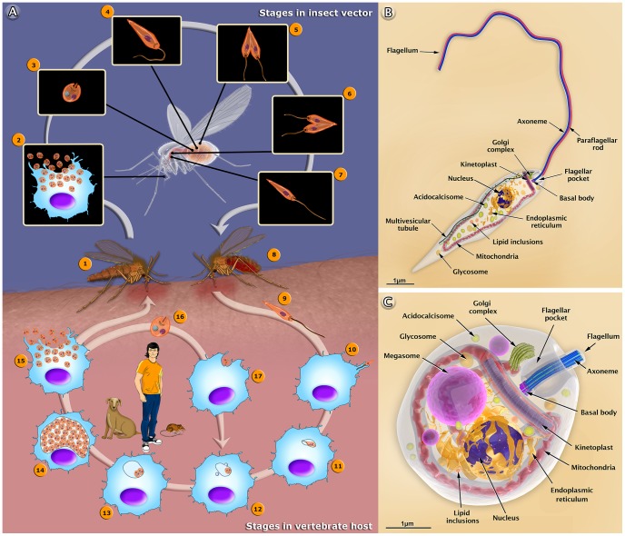
(A) The female sandfly (1) insect bites an infected mammal during the blood meal. Infected macrophages (2) with amastigote forms. (3) Amastigote form. (4) Amastigotes transform into procyclic promastigotes. (5) Procyclic promastigotes multiply in the midgut. (6) Promastigotes migrate toward the stomodeal valve in the anterior midgut and re-initiate cell division. (7) Promastigotes transform into infective metacyclic promastigotes. (8) The female sandfly releases the metacyclic promastigotes into a new mammalian host via regurgitation during the blood meal. (9) Metacyclic promastigotes. (10) Metacyclic promastigotes infect macrophages. (11) Metacyclic promastigotes transform into amastigotes. (12) Amastigotes attach to the membrane of the parasitophorous vacuole. (13) Amastigotes multiply in the vacuole. (14) Intense amastigote multiplication. (15) Amastigotes burst out of the cell. (16) Amastigote form. (17) An amastigote infects a macrophage. In the central portion of the figure, we added the most important reservoirs involved in the maintenance of the parasite. Schematic 3D representations of the organelles found in the Leishmania promastigote (B) and amastigote (C).
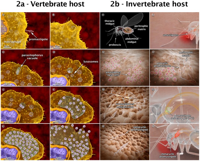
(A) Attachment of a promastigote to the macrophage surface. (B) The process of internalization via phagocytosis begins with the formation of pseudopods (C), leading to the formation of the parasitophorous vacuole (PV). In the PV, the promastigote transforms into an amastigote. (D) Recruitment and fusion of host cell lysosomes with the PV takes place. (E) In the PV, amastigotes divide several times. (F–G) Intense multiplication generates several hundreds of amastigotes. (H) The host cell bursts, and the parasites reach the extracellular space. (I) Schematic view of female sandfly showing the digestive tract. (J) During a blood meal, a female sandfly ingests infected macrophages with amastigote forms present in the blood of the vertebrate host. (K) Amastigotes form “nest cells” in the abdominal midgut. (L) Amastigotes transform into procyclic promastigotes. (M) Promastigotes multiply and attach to the midgut epithelium. (N) Parasites migrate toward the anterior midgut, resume replication and start to produce promastigote secretory gel (PSG). (O) Promastigotes transform into infective metacyclic promastigotes. (P) Metacyclic promastigotes infect a new mammalian host via regurgitation during the blood meal. These images are based on micrographs obtained by scanning and transmission electron microscopy and by video microscopy.
References
-
- Alvar J, Vélez ID, Bern C, Herrero M, Desjeux P, et al. (2012) Leishmaniasis worldwide and global estimates of its incidence. PLoS One 7: e35671 doi: 10.1371/journal.pone.0035671 - DOI - PMC - PubMed
-
- Pollock E, Chandler P, Sweller J (2002) Assimilating complex information. Learn Instruct 12: 61–86.
-
- Tversky B, Morrison JB (2002) Animation: can it facilitate? Int J Hum Comput Stud 57: 247–262.
-
- Ribeiro JM, Rossignol PA, Spielman A (1986) Blood finding strategy of a capillary - feeding sandfly Lutzomyia longipalpis . Comp Biochem Physiol 83: 683–6. - PubMed
Publication types
MeSH terms
LinkOut - more resources
Full Text Sources
Other Literature Sources
Medical
