. 2013;9(10):e1003657.
doi: 10.1371/journal.ppat.1003657. Epub 2013 Oct 10.

New world bats harbor diverse influenza A viruses

Affiliations

New world bats harbor diverse influenza A viruses

Suxiang Tong et al. PLoS Pathog. 2013.

Abstract

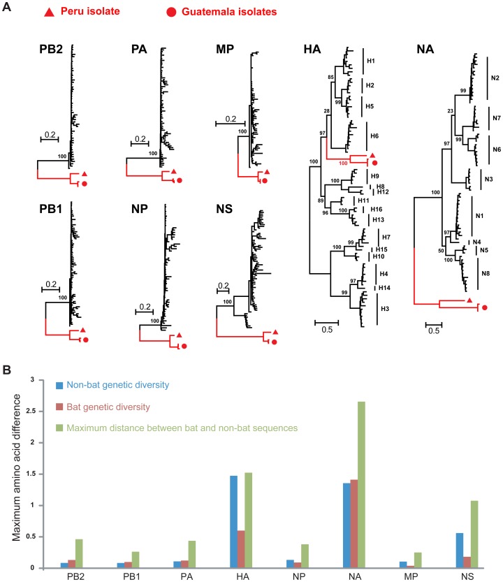
Aquatic birds harbor diverse influenza A viruses and are a major viral reservoir in nature. The recent discovery of influenza viruses of a new H17N10 subtype in Central American fruit bats suggests that other New World species may similarly carry divergent influenza viruses. Using consensus degenerate RT-PCR, we identified a novel influenza A virus, designated as H18N11, in a flat-faced fruit bat (Artibeus planirostris) from Peru. Serologic studies with the recombinant H18 protein indicated that several Peruvian bat species were infected by this virus. Phylogenetic analyses demonstrate that, in some gene segments, New World bats harbor more influenza virus genetic diversity than all other mammalian and avian species combined, indicative of a long-standing host-virus association. Structural and functional analyses of the hemagglutinin and neuraminidase indicate that sialic acid is not a ligand for virus attachment nor a substrate for release, suggesting a unique mode of influenza A virus attachment and activation of membrane fusion for entry into host cells. Taken together, these findings indicate that bats constitute a potentially important and likely ancient reservoir for a diverse pool of influenza viruses.


Conflict of interest statement

The authors have declared that no competing interests exist.

Figures

Figure 1
Figure 1. Evolution of influenza virus in New World bats.
(a) Phylogenetic relationships of influenza A viruses sampled from bats (red branches) and other animals (‘non-bat’, black branches) based on the amino acid sequences of each gene segment. (b) The maximum amino acid distances are shown within and between the bat and non-bat sequences, and colored accordingly. These levels of diversity also effectively act as a scale-bar for the trees shown above.
Figure 2
Figure 2. Crystal structures of A/bat/Peru/10 HA.
(A) Overall structure of A/bat/Peru/10 HA. The H18 HA trimer consists of three identical monomers with one RBS per monomer. HA1 is highlighted in green and HA2 in cyan. N-linked glycans observed in the electron density maps are shown with yellow carbons. (B) The A/bat/Peru/10 HA putative RBS (in crystal 1, Table S6) in ribbon representation with the side chains of key binding residues shown. The three highly conserved residues (W153, H183, and Y195) in HAs are colored with green carbon atoms, whereas nine residues that are conserved in A/bat/Peru/10 H18 and two H17 HAs from bat influenza viruses A/little yellow-shouldered bat/Guatemala/164/2009 (H17N10) (GU09-164) and A/little yellow-shouldered bat/Guatemala/060/2010 (H17N10) (GU10-060) are labeled in red. E190 and G225 are also conserved, especially in avian influenza A viruses. (C) Molecular surface of the putative RBS of A/bat/Peru/10 HA compared to the RBS of 2009 H1 HA from A/California/04/2009 (H1N1) (PDB code 3UBQ). A canonical sialic acid is modeled in the HA for comparison as observed in other HA structures. The RBS of A/bat/Peru/10 HA is shallower and wider than 2009 H1 HA with no space for the glycerol moiety of sialic acid (indicated by the red arrow). For comparison, figures (B) and (C) are generated in the same orientation.
Figure 3
Figure 3. SDS-PAGE of A/bat/Peru/10 HA0 (lanes 1 to 4) and its mature HA (lanes 5 to 8) in trypsin susceptibility assay.
A/bat/Peru/10 HA with a monobasic cleavage site was expressed in its HA0 form in a baculovirus expression system. Lanes 1 and 2 show A/bat/Peru/10 native HA0 at pH 8.0 and pH 4.9, respectively, while lanes 3 and 4 show the equivalent reducing gel of HA0 treated with trypsin at pH 8.0 and pH 4.9, respectively. Similarly, lanes 5 and 6 show A/bat/Peru/10 mature HA at pH 8.0 and pH 4.9, respectively, while lanes 7 and 8 show the equivalent reducing gel of mature HA treated with trypsin at pH 8.0 and pH 4.9, respectively.
Figure 4
Figure 4. Crystal structures of A/bat/Peru/10 NAL.
(A) Overall structure of A/bat/Peru/10 NAL with a conserved calcium binding site. The N11 NAL tetramer is viewed from above the viral surface, and consists of four identical monomers with C4 symmetry. One monomer is colored in six different colors to illustrate the canonical β-propeller shape of six four-stranded, anti-parallel β-sheets. The putative active site is located on the membrane-distal surface (on top of the molecule). The observed N-linked glycosylation sites are shown with attached carbohydrates. A single calcium ion is shown in red spheres. (B) The A/bat/Peru/10 NAL putative active site (crystal form 1, Table S6) with the conserved catalytic and active site residues in other NAs shown as well as other polar and charged residues. The six residues (R118, W178, S179, R224, E276 and E425) conserved in all influenza NAs are colored with green carbon atoms in contrast to other putative active site residues in yellow carbon atoms, whereas eight residues that are conserved in two bat influenza N10 and N11 NAL proteins are labeled in red. (C) Molecular surface of the active site of A/bat/Peru/10 N11 NAL and 1918 N1 NA from A/Brevig Mission/1/18 (H1N1) (PDB code 3BEQ). A canonical sialic acid is modeled in A/bat/Peru/10 NAL as in other NA structures and appears to collide with the NA putative active site around the glycerol moiety (as indicated by the red arrow). The putative active site pocket of A/bat/Peru/10 NAL is much wider than 1918 N1 NA. For comparison, figures (B) and (C) are generated in the same orientation.

References

    1. Garten RJ, Davis CT, Russell CA, Shu B, Lindstrom S, et al. (2009) Antigenic and genetic characteristics of swine-origin 2009 A(H1N1) influenza viruses circulating in humans. Science 325: 197–201. - PMC - PubMed
    1. Tong S, Li Y, Rivailler P, Conrardy C, Castillo DA, et al. (2012) A distinct lineage of influenza A virus from bats. Proc Natl Acad Sci USA 109: 4269–4274. - PMC - PubMed
    1. Li W, Shi Z, Yu M, Ren W, Smith C, et al. (2005) Bats are natural reservoirs of SARS-like coronaviruses. Science 310: 676–679. - PubMed
    1. Tong S, Conrardy C, Ruone S, Kuzmin IV, Guo X, et al. (2009) Detection of novel SARS-like and other coronaviruses in bats from Kenya. Emerg Infect Dis 15: 482–485. - PMC - PubMed
    1. Turmelle AS, Olival KJ (2009) Correlates of viral richness in bats (order Chiroptera). Ecohealth 6: 522–539. - PMC - PubMed

Publication types

MeSH terms

Substances