. 2013;9(10):e1003708.
doi: 10.1371/journal.ppat.1003708. Epub 2013 Oct 10.

Regulation of innate responses during pre-patent schistosome infection provides an immune environment permissive for parasite development

Affiliations

Regulation of innate responses during pre-patent schistosome infection provides an immune environment permissive for parasite development

Diana K Riner et al. PLoS Pathog. 2013.

Abstract

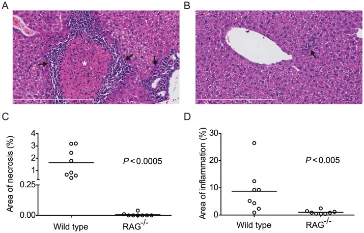
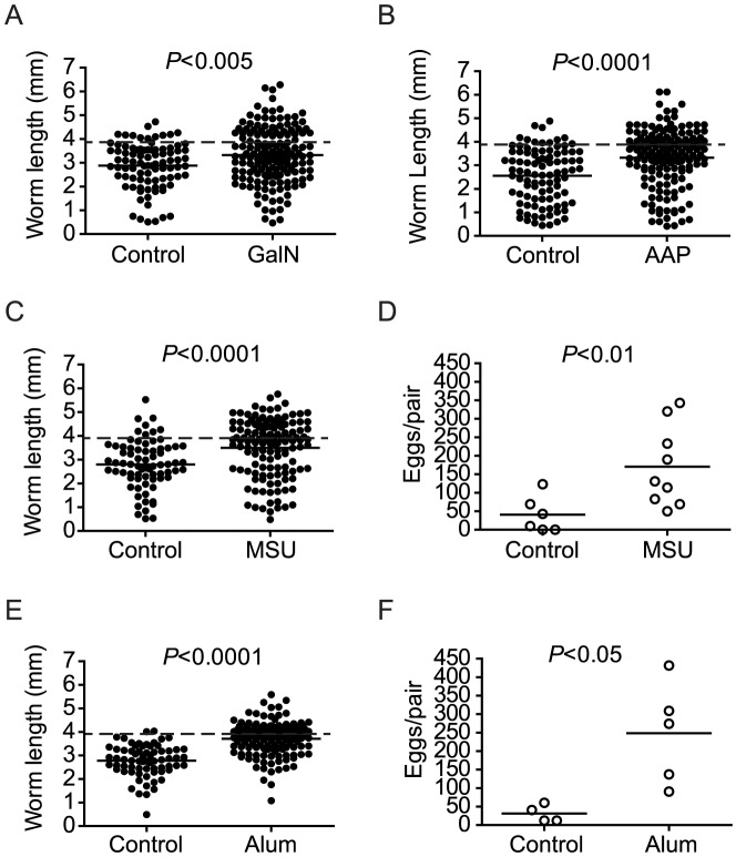
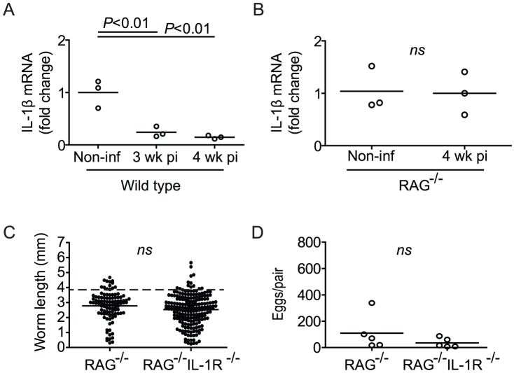
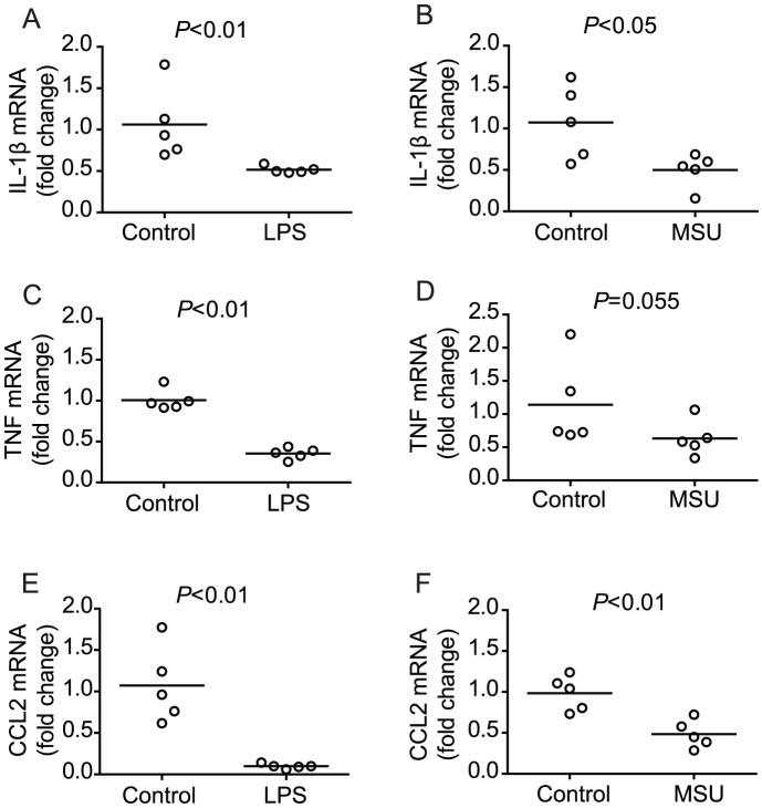
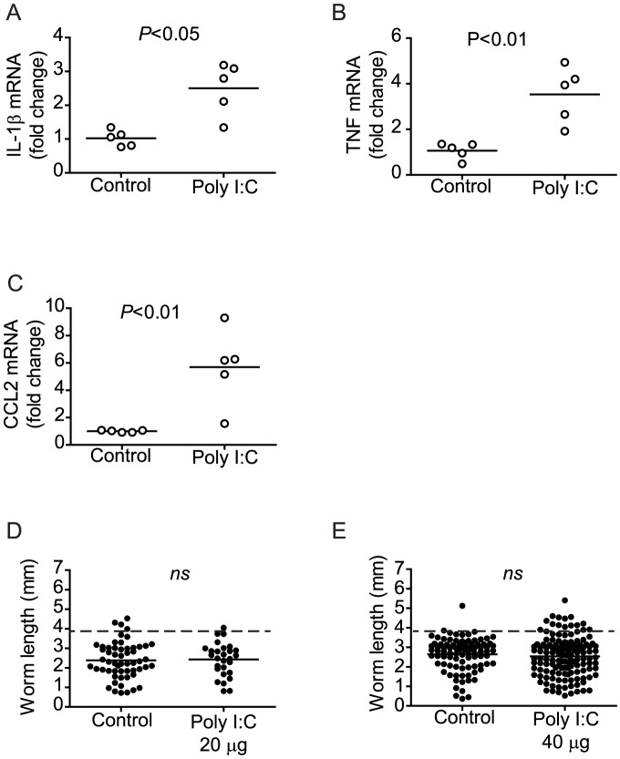
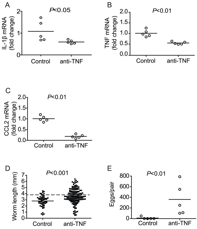
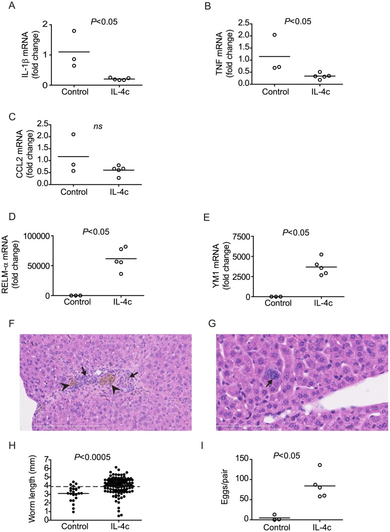
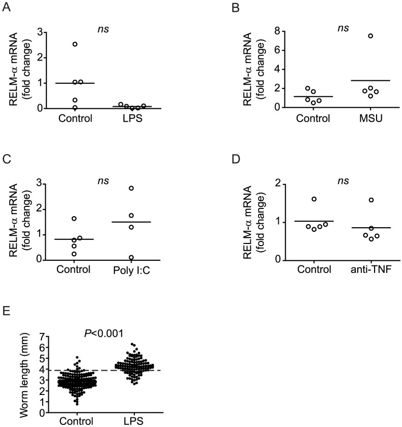
Blood flukes of the genus Schistosoma infect over 200 million people, causing granulomatous pathology with accompanying morbidity and mortality. As a consequence of extensive host-parasite co-evolution, schistosomes exhibit a complex relationship with their hosts, in which immunological factors are intimately linked with parasite development. Schistosomes fail to develop normally in immunodeficient mice, an outcome specifically dependent on the absence of CD4⁺ T cells. The role of CD4⁺ T cells in parasite development is indirect and mediated by interaction with innate cells, as repeated toll-like receptor 4 stimulation is sufficient to restore parasite development in immunodeficient mice in the absence of CD4⁺ T cells. Here we show that repeated stimulation of innate immunity by an endogenous danger signal can also restore parasite development and that both these stimuli, when administered repeatedly, lead to the regulation of innate responses. Supporting a role for regulation of innate responses in parasite development, we show that regulation of inflammation by neutralizing anti-TNF antibodies also restores parasite development in immunodeficient mice. Finally, we show that administration of IL-4 to immunodeficient mice to regulate inflammation by induction of type 2 responses also restores parasite development. These findings suggest that the type 2 response driven by CD4⁺ T cells during pre-patent infection of immunocompetent hosts is exploited by schistosomes to complete their development to reproductively mature adult parasites.


Conflict of interest statement

The authors have declared that no competing interests exist.

Figures

Figure 1
Figure 1. Pre-patent S. mansoni infection does not induce liver necrosis and inflammation in RAG−/− mice.
Wild type (C57BL/6) and RAG−/− mice were infected with S. mansoni cercariae via percutaneous tail exposure and livers were removed for histological analysis at 4 weeks p.i. (A) Representative H&E-stained liver section from a wild type mouse, exhibiting inflammatory cell infiltration (arrows) in periportal area and surrounding a focus of coagulative necrosis (⋆). (B) Representative H&E-stained liver section from a RAG−/− mouse. Note small and isolated cluster of inflammatory cells (arrow). (C) Average percent area of tissue section occupied by coagulative necrosis was calculated for each animal (n = 8, pooled from two independent experiments). Horizontal bars represent mean values for each experimental group. (D) Average percent area of tissue section occupied by inflammatory infiltrate was calculated for each animal (n = 8, pooled from two independent experiments). Horizontal bars represent mean values for each experimental group. P values were determined using the Mann Whitney test. Scale bars in A and B are 300 µm in length.
Figure 2
Figure 2. Chronic hepatocyte injury or DAMP administration facilitates S. mansoni development in RAG−/− mice.
Groups of RAG−/− mice were infected with S. mansoni and treated throughout pre-patent infection with the indicated hepatotoxins or inflammatory stimuli, as described in Materials and Methods. Parasites were perfused from the portal tract and livers removed for egg enumeration at 6 weeks p.i. (A) Length of male worms recovered from RAG−/− mice chronically treated with D-(+)-galactosamine hydrochloride (GalN). (B) Length of male worms recovered from RAG−/− mice chronically treated with acetaminophen (AAP). (C and E) Length of male worms and (D and F) liver egg burdens from RAG−/− mice chronically treated with monosodium urate (MSU; C and D) or alum (E and F). Horizontal bars represent mean values for each experimental group. Groups of 4 to 5 mice were used for each experimental condition. P values for worm lengths determined using student's T-test with Welch's correction. P values for egg production determined using the Mann Whitney test. Data shown for the MSU experiment are pooled from two independent experiments. Dashed lines in A, B, C, and E indicate the average length of male S. mansoni worms recovered from wild type mice at day 42 post infection (3.9 mm).
Figure 3
Figure 3. Impaired S. mansoni development in RAG−/− mice correlates with maintenance of IL-1β transcription.
(A and B) Groups of wild type (A) and RAG−/− mice (B) were infected percutaneously with S. mansoni cercariae, euthanized at the indicated time points and spleens removed for analysis of IL-1β mRNA levels by real-time PCR. Horizontal bars represent mean values for each experimental group. For wild type mice (A), P values were calculated by ANOVA with Tukey's post-test. For RAG−/− mice (B), P value was calculated using the Mann Whitney test. (C) Length of male worms and (D) liver egg burdens from S. mansoni-infected RAG−/− and RAG−/− IL-1R−/− mice at 6 weeks p.i. Horizontal bars represent mean values for each experimental group. P values were determined using student's T-test with Welch's correction and the Mann Whitney test. Dashed line in C indicates the average length of male S. mansoni worms recovered from wild type mice at day 42 post infection (3.9 mm).
Figure 4
Figure 4. Chronic LPS or MSU administration restores regulation of pro-inflammatory cytokine transcription in infected RAG−/− mice.
Groups of S. mansoni-infected RAG−/− mice were repeatedly treated throughout pre-patent infection with either LPS (A, C, E) or MSU (B, D, F), as described in Materials and Methods. At 6 weeks p.i., mice were euthanized and spleens removed for analysis of IL-1β (A and B), TNF (C and D) and CCL2 (E and F) mRNA content by real-time PCR. Horizontal bars represent mean values for each experimental group. P values were calculated using the Mann Whitney test.
Figure 5
Figure 5. Chronic poly I:C administration does not restore regulation of pro-inflammatory cytokine transcription or parasite development in infected RAG−/− mice.
Groups of S. mansoni-infected RAG−/− mice were repeatedly treated throughout pre-patent infection with poly I:C, as described in Materials and Methods. At 6 weeks p.i., mice were euthanized, parasites perfused from the portal tract and spleens removed for analysis of IL-1β (A), TNF (B) and CCL2 (C) mRNA content by real-time PCR. (D and E) Length of male worms recovered from RAG−/− mice treated repeatedly with 20 µg (D) or 40 µg (E) doses of poly I:C. Horizontal bars represent mean values for each experimental group. P values were calculated using the Mann Whitney test and student's T-test with Welch's correction. Dashed lines in D and E indicate the average length of male S. mansoni worms recovered from wild type mice at day 42 post infection (3.9 mm).
Figure 6
Figure 6. Chronic administration of anti-TNF antibody restores regulation of pro-inflammatory cytokine transcription and parasite development in infected RAG−/− mice.
Groups of S. mansoni-infected RAG−/− mice were repeatedly treated throughout pre-patent infection with anti-TNF antibody, as described in Materials and Methods. At 6 weeks p.i., mice were euthanized, parasites perfused from the portal tract and spleens removed for analysis of IL-1β (A), TNF (B) and CCL2 (C) mRNA content by real-time PCR. (D) Length of male worms and (E) liver egg burdens from RAG−/− mice treated with anti-TNF antibody. Horizontal bars represent mean values for each experimental group. P values were calculated using the Mann Whitney test and student's T-test with Welch's correction. Dashed line in D indicates the average length of male S. mansoni worms recovered from wild type mice at day 42 post infection (3.9 mm).
Figure 7
Figure 7. Chronic administration of IL-4 restores regulation of pro-inflammatory cytokine transcription and parasite development in infected RAG−/− mice.
Groups of S. mansoni-infected RAG−/− mice were repeatedly treated throughout pre-patent infection with IL-4 complex (IL-4c), as described in Materials and Methods. At 6 weeks p.i., mice were euthanized, parasites perfused from the portal tract and spleens (A–C) and livers (D, E) removed for analysis of IL-1β (A), TNF (B), CCL2 (C), RELM-α (D) and YM1 (E) mRNA content by real-time PCR. (F and G) Representative low- (F; 20×) and high-power (G; 40×) fields of H&E-stained liver sections from infected RAG−/− mice treated with IL-4c. In (F), note inflammatory infiltrate (arrows), composed partly of mononuclear phagocytes containing brown by-product of schistosome hemoglobin degradation (arrowheads). In (G), arrow indicates a multinucleate giant cell. Lengths of scale bars are 300 µm (F) and 100 µm (G). (H) Length of male worms and (I) liver egg burdens from RAG−/− mice treated with IL-4. Horizontal bars represent mean values for each experimental group. P values were calculated using the Mann Whitney test and student's T-test with Welch's correction. Dashed line in H indicates the average length of male S. mansoni worms recovered from wild type mice at day 42 post infection (3.9 mm).
Figure 8
Figure 8. Restoration of parasite development is not dependent on M2 responses.
Groups of S. mansoni-infected RAG−/− mice were repeatedly treated throughout pre-patent infection with either LPS (A) or MSU (B), poly I:C (C) or anti-TNF antibody (D) as described in Materials and Methods. At 6 weeks p.i., mice were euthanized and livers removed for analysis of RELM-α mRNA content by real-time PCR. E, groups of RAG−/−c −/− were infected with S. mansoni and repeatedly treated throughout per-patent infection with LPS or PBS (control), as described in Materials and Methods. Parasites were perfused from the portal system at 6 weeks p.i. and the length of the male worms measured. Horizontal bars represent mean values for each experimental group. P values were calculated using the Mann Whitney test. Dashed line in E indicates the average length of male S. mansoni worms recovered from wild type mice at day 42 post infection (3.9 mm).

References

    1. Davies SJ, Grogan JL, Blank RB, Lim KC, Locksley RM, et al. (2001) Modulation of Blood Fluke Development in the Liver by Hepatic CD4+ Lymphocytes. Science 294: 1358–1361. - PubMed
    1. Lamb EW, Crow ET, Lim KC, Liang YS, Lewis FA, et al. (2007) Conservation of CD4+ T cell-dependent developmental mechanisms in the blood fluke pathogens of humans. Int J Parasitol 37: 405–415. - PMC - PubMed
    1. Harrison RA, Doenhoff MJ (1983) Retarded development of Schistosoma mansoni in immunosuppressed mice. Parasitology 86: 429–438. - PubMed
    1. Lamb EW, Walls CD, Pesce JT, Riner DK, Maynard SK, et al. (2010) Blood fluke exploitation of non-cognate CD4+ T cell help to facilitate parasite development. PLoS Pathog 6: e1000892. - PMC - PubMed
    1. Stadecker MJ, Asahi H, Finger E, Hernandez HJ, Rutitzky LI, et al. (2004) The immunobiology of Th1 polarization in high-pathology schistosomiasis. Immunol Rev 201: 168–179. - PubMed

Publication types

MeSH terms