. 2013 Oct 11:4:706.
doi: 10.3389/fpsyg.2013.00706. eCollection 2013.

Separating lexical-semantic access from other mnemonic processes in picture-name verification

Affiliations

Separating lexical-semantic access from other mnemonic processes in picture-name verification

Jason F Smith et al. Front Psychol. .

Abstract

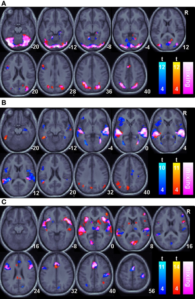
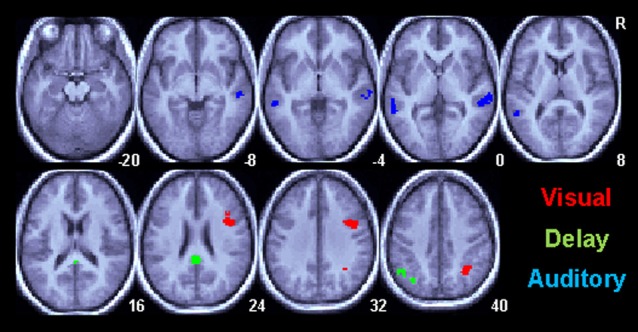
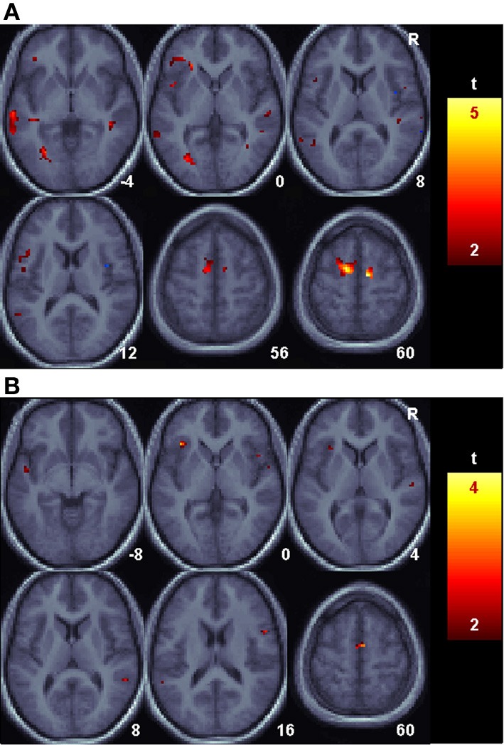
We present a novel paradigm to identify shared and unique brain regions underlying non-semantic, non-phonological, abstract, audio-visual (AV) memory vs. naming using a longitudinal functional magnetic resonance imaging experiment. Participants were trained to associate novel AV stimulus pairs containing hidden linguistic content. Half of the stimulus pairs were distorted images of animals and sine-wave speech versions of the animal's name. Images and sounds were distorted in such a way as to make their linguistic content easily recognizable only after being made aware of its existence. Memory for the pairings was tested by presenting an AV pair and asking participants to verify if the two stimuli formed a learned pairing. After memory testing, the hidden linguistic content was revealed and participants were tested again on their recollection of the pairings in this linguistically informed state. Once informed, the AV verification task could be performed by naming the picture. There was substantial overlap between the regions involved in recognition of non-linguistic sensory memory and naming, suggesting a strong relation between them. Contrasts between sessions identified left angular gyrus and middle temporal gyrus as key additional players in the naming network. Left inferior frontal regions participated in both naming and non-linguistic AV memory suggesting the region is responsible for AV memory independent of phonological content contrary to previous proposals. Functional connectivity between angular gyrus and left inferior frontal gyrus and left middle temporal gyrus increased when performing the AV task as naming. The results are consistent with the hypothesis that, at the spatial resolution of fMRI, the regions that facilitate non-linguistic AV associations are a subset of those that facilitate naming though reorganized into distinct networks.

Keywords: crossmodal; fMRI; language; memory.


Figures

Figure 1
Figure 1
Example experimental visual stimuli. Two example visual stimuli from the experiment are shown. The images to the left are actual stimuli used in the experiment. The images to the right are the unfiltered images from which the experimental stimuli were created. All scanning trials used the filtered (left) stimuli. Fractal images were used with permission from www.fractalrecursions.com.
Figure 2
Figure 2
Task related BOLD increases for correct trials relative to an arbitrary resting baseline for all sessions. Only areas surviving a p < 0.05(corrected) cluster level threshold are shown on the mean normalized anatomical image from all twelve subjects. The Non-linguistic session (N) is shown in a blue-cyan scale and the Informed, linguistic session (L) is shown in red-yellow scale. Overlapping areas are indicated by color mixing according to the color scale as shown in the legend. (A). The Visual cue portion of the task. (B). The Delay period portion of the task. (C). The Auditory target portion of the task.
Figure 3
Figure 3
Task related BOLD increases for correct trials in the Informed/Linguistic session relative to the non-linguistic session for all subcomponents of the task. Only areas surviving a p < 0.05(corrected) cluster level threshold are shown on the mean normalized anatomical image from all twelve subjects. The Visual cue (V) period is shown in red, the Delay (D) period is green and the Auditory (A) target period is shown in red.
Figure 4
Figure 4
Language related changes in functional connectivity with the angular gyrus. Shown are areas with reliably greater BOLD signal relative to baseline for the Uninformed, non-linguistic session that nonetheless had reliably greater functional connectivity with the angular gyrus during the Informed, linguistic session. (A). The Delay period portion of the task. (B). The Auditory target portion of the task.

References

    1. Andrews T. J., Schluppeck D. (2003). Neural responses to Mooney images reveal a modular representation of faces in human visual cortex. Neuroimage 21, 91–98 10.1016/j.neuroimage.2003.08.023 - DOI - PubMed
    1. Bak T. H., Hodges J. R. (2004). The effects of motor neurone disease on language: further evidence. Brain Lang. 89, 354–361 10.1016/S0093-934X(03)00357-2 - DOI - PubMed
    1. Barde L. H., Thompson-Schill S. L. (2002). Models of functional organization of the lateral prefrontal cortex in verbal working memory: evidence in favor of the process model. J. Cogn. Neurosci. 14, 1054–1063 10.1162/089892902320474508 - DOI - PubMed
    1. Beauchamp M. S., Lee K. E., Argall B. D., Martin A. (2004). Integration of auditory and visual information about objects in superior temporal sulcus. Neuron 41 809–823 10.1016/S0896-6273(04)00070-4 - DOI - PubMed
    1. Beckmann C. F., Jenkinson M., Smith S. M. (2003). General multilevel linear modeling for group analysis in FMRI. Neuroimage 20, 1052–1063 10.1016/S1053-8119(03)00435-X - DOI - PubMed

LinkOut - more resources