. 2013 Oct 10;8(10):e75443.
doi: 10.1371/journal.pone.0075443. eCollection 2013.

Targeting fungal genes by diced siRNAs: a rapid tool to decipher gene function in Aspergillus nidulans

Affiliations

Targeting fungal genes by diced siRNAs: a rapid tool to decipher gene function in Aspergillus nidulans

Natarajaswamy Kalleda et al. PLoS One. .

Abstract

Background: Gene silencing triggered by chemically synthesized small interfering RNAs (siRNAs) has become a powerful tool for deciphering gene function in many eukaryotes. However, prediction and validation of a single siRNA duplex specific to a target gene is often ineffective. RNA interference (RNAi) with synthetic siRNA suffers from lower silencing efficacy, off-target effects and is cost-intensive, especially for functional genomic studies. With the explosion of fungal genomic information, there is an increasing need to analyze gene function in a rapid manner. Therefore, studies were performed in order to investigate the efficacy of gene silencing induced by RNase III-diced-siRNAs (d-siRNA) in model filamentous fungus, Aspergillus nidulans.

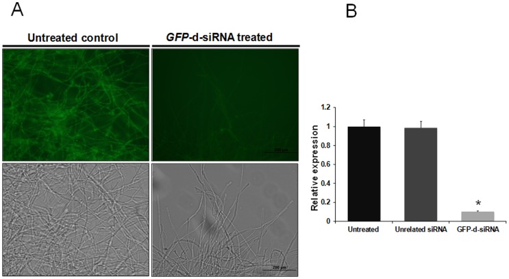
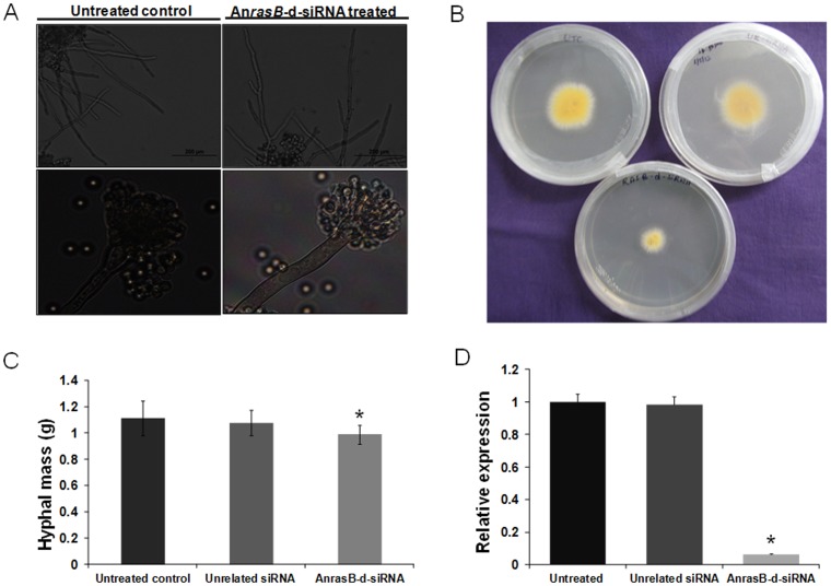
Methodology/principal findings: Stable expression of heterologous reporter gene in A. nidulans eases the examination of a new RNAi-induction route. Hence, we have optimized Agrobacterium tumefaciens-mediated transformation (AMT) of A. nidulans for stable expression of sGFP gene. This study demonstrates that the reporter GFP gene stably introduced into A. nidulans can be effectively silenced by treatment of GFP-d-siRNAs. We have shown the down-regulation of two endogenous genes, AnrasA and AnrasB of A. nidulans by d-siRNAs. We have also elucidated the function of an uncharacterized Ras homolog, rasB gene, which was found to be involved in hyphal growth and development. Further, silencing potency of d-siRNA was higher as compared to synthetic siRNA duplex, targeting AnrasA. Silencing was shown to be sequence-specific, since expression profiles of other closely related Ras family genes in d-siRNA treated AnrasA and AnrasB silenced lines exhibited no change in gene expression.

Conclusions/significance: We have developed and applied a fast, specific and efficient gene silencing approach for elucidating gene function in A. nidulans using d-siRNAs. We have also optimized an efficient AMT in A. nidulans, which is useful for stable integration of transgenes.


Conflict of interest statement

Competing Interests: The authors have declared that no competing interests exist.

Figures

Figure 1
Figure 1. T-DNA map of sGFP expression vector.
sGFP gene was cloned under the control of Aspergillus nidulans TrpC promoter and marker gene (HPT) was fused to CaMV35S promoter in the T-DNA back bone of pCAMBIA1300 binary vector.
Figure 2
Figure 2. Molecular characterization of A. nidulans sGFP transformants.
(A) DNA from several independent hygromycin resistant A. nidulans mycelia was extracted and the presence of sGFP gene (200 bp amplicon) was evaluated by PCR amplification. M. Ladder; PC. pCAMBIA 1300-sGFP plasmid control; 1–4. A. nidulans transformants represented as AnGFP 101, AnGFP 102, AnGFP 103, AnGFP 104; UT. Untransformed control. (B) DNA from several independent hygromycin resistant A. nidulans mycelia was extracted and the presence of HPT gene was evaluated by PCR amplification. M. Ladder; UT. Untransformed control; 1–4. AnGFP 101, AnGFP 102, AnGFP 103, AnGFP 104 fungal transformants. (C) Southern hybridization was performed using genomic DNA extracted from fungal transformants, DNA was (10 µg) digested with XhoI and probed with radio labelled HPT probe. UT. Untransformed control; 1–4. AnGFP 101, AnGFP 102, AnGFP 103, AnGFP 104 fungal transformants. (D) Semi-quantitative RT-PCR analysis for evaluation of expression of sGFP in A. nidulans transformants. Upper panel indicates sGFP expression, 1–4. AnGFP 101, AnGFP 102, AnGFP 103, AnGFP 104 fungal transformants; UT. Untransformed control. Lower panel: normalization of gene expression by AnActin as internal control.
Figure 3
Figure 3. Detection of GFP in different developmental stages of A. nidulans (AnGFP 101) transformant.
GFP expression was examined using an OLYMPUS BX-51 fluorescent microscope under UV light (right) or white light conditions (left) in fungal spores, mycelia and conidiophores. Expression of GFP in AnGFP 101 transformant (left panel) showed strong GFP fluorescence in all the developmental stages, whereas GFP expression was completely absent in untransformed control AnWT (right panel). All the comparative pictures in AnGFP 101 and AnWT were taken at same magnifications using fluorescent microscopy.
Figure 4
Figure 4. Strategies for generation of dsRNAs and d-siRNAs.
(A) PCR template strategy was utilized for obtaining sGFP, AnrasA and AnrasB dsRNA in a single T7 transcription reaction. T7 promoter sequence was added to both forward and reverse gene specific primers of sGFP, AnrasA and AnrasB, and then PCR amplification was performed in order to generate templates for dsRNA synthesis. (B) For generation of template DNA for unrelated MiAchE, PCR amplification was performed on pGEM T-MiAchE vector using M13 primers. This amplification includes the T7 promoter to one end and SP6 promoter to another end of MiAchE. (C) PCR templates for transcription of respective target genes. M. ladder; B. blank; sGFP, AnrasA, AnrasB and MiAchE target gene templates. (D) Synthesis of dsRNAs with T7 or SP6 in vitro transcription reactions. M. Ladder; sGFP, AnrasA, AnrasB and MiAchE dsRNAs. (E) 20% PAGE analysis of purified diced siRNAs. M-Ladder, sGFP, AnrasA, AnrasB and MiAchE d-siRNAs. The d-siRNAs of all target genes were generated by cleaving respective dsRNAs with RNase III at 37°C for 30 mins, followed by subsequent purifications and finally dissolved in nuclease free water.
Figure 5
Figure 5. Evaluation of GFP silencing by GFP-d-siRNAs in A. nidulans.
(A) Evaluation of green fluorescence in GFP-d-siRNA treated mycelia. A. nidulans (1×10⁻⁴/ ml) spores were added to 24 well culture plate and germination was induced at 37°C for 6 h, subsequently 25 nM final concentration of GFP-d-siRNAs were added and treated for another 12 h. Pictures by microscopy were taken 18 h after seeding. The (left panel) untreated fungal mycelia was showing brighter GFP fluorescence, whereas (right panel) the treatment of fungal mycelia with 25 nM final concentration GFP-d-siRNA resulted in drastic reduction in GFP fluorescence. Upper panel showing the microscopic pictures under the UV light and lower panel showing the microscopic pictures under the white light. (B) Quantification of GFP silencing by qRT-PCR. Relative GFP expression was measured in untreated control, unrelated siRNA treated and GFP-d-siRNA treated mycelia using comparative D cycle threshold (CT) method. GFP values were normalized to AnActin values. The data represent the means of three replicates. Values were compared using t test. * Significant difference at P<0.05 as compared to untreated control.
Figure 6
Figure 6. Evaluation of AnrasA silencing by AnrasA-d-siRNA in A. nidulans.
(A) Determination of phenotypic changes in A. nidulans. The germinating conidia were treated with AnrasA-d-siRNA for 12 h, then light microscopic analysis (18 h after seeding) revealed that the majority of treated conidia were swollen but unable to form hyphal tissue compared to control. (B) Radial growth assay. Germinating conidia were treated with AnrasA-d-siRNA and spotted onto center of the ACM agar plate and radial outgrowth was monitored 48 h post-inoculation. Radial growth was drastically reduced in AnrasA silenced lines compared to controls. (C) Comparison of the total biomass. Germinating conidia were treated with AnrasA-d-siRNA in ACM broth for 48 h and total fresh biomass formation was determined. The data represent the means of three replicates. Values were compared using t test. * Significant difference at P<0.05 as compared to untreated control. (D) Quantification of AnrasA silencing by qRT-PCR. Relative AnrasA expression was measured in untreated control, unrelated siRNA treated and AnrasA-d-siRNA treated mycelia using comparative D cycle threshold (CT) method. AnrasA values were normalized to AnActin values. The data represent the means of three replicates. Values were compared using t test. * Significant difference at P<0.05 as compared to untreated control.
Figure 7
Figure 7. Evaluation of AnrasB down-regulation by AnrasB-d-siRNA in A. nidulans.
(A) Assessment of phenotypic changes in A. nidulans. In upper panel germinating conidia were treated with AnrasB-d-siRNA for 12 h, and then light microscopic analysis was performed. The AnrasB silenced lines possess the irregular apical branching when compared to control. In the lower panel, fungal tissue was collected from ACM solid agar media containing AnrasB silenced lines and control lines, then viewed using OLYMPUS BX-51 microscope under the white light conditions. (B) Radial mycelial growth assay. Germinating conidia were treated with AnrasB-d-siRNA and spotted onto center of the ACM agar plate and radial outgrowth of mycelia was monitored 48 h post-inoculation. Radial growth was significantly reduced in AnrasB silenced lines compared to controls. (C) Determination of the total biomass. Germinating conidia were treated with AnrasB-d-siRNA in ACM broth for 48 h and total fresh biomass formation was determined. The data represent the means of three replicates. Values were compared using t test. * Significant difference at P<0.05 as compared to untreated control. (D) Quantification of AnrasB silencing by qRT-PCR. Relative AnrasB expression was measured in AnrasB-d-siRNA treated mycelia and controls using comparative D cycle threshold (CT) method. AnrasB values were normalized to AnActin values. The data represent the means of three replicates. Values were compared using t test. * Significant difference at P<0.05 as compared to untreated control.
Figure 8
Figure 8. Determination of silencing efficacies of synthetic siRNA and d-siRNA.
Real-Time PCR analysis was performed to compare the silencing potency of chemically synthesized siRNA and RNase III-diced-siRNA targeting rasA endogenous gene in A. nidulans. A. nidulans spores were germinated for 6 h in ACM medium. Subsequently, 25 nM final concentration of synthetic AnrasA siRNA (rasA-c-siRNA) and RNase III generated AnrasA siRNA (rasA-d-siRNA) were added and incubated for another 12 h. Then mycelial tissues were collected and RNA was isolated. Relative AnrasA expression was measured in untreated control, unrelated siRNA treated, rasA-d-siRNA treated and rasA-c-siRNA treated mycelia using comparative D cycle threshold (CT) method. AnrasA values were normalized to AnActin values. The data represent the means of three replicates. Values were compared using t test. * Significant difference at P<0.05 as compared to untreated control.
Figure 9
Figure 9. Expression profiles of Ras family genes in d-siRNA treated lines.
(A) Estimation of relative expression levels of Ras family genes in untreated control and AnrasA-d-siRNA treated A. nidulans. Relative quantification was performed by qRT-PCR using comparative D cycle threshold (CT) method. Expression levels of AnrasB, AnrhB, An4873, AnmedA and An7661 in untreated control was showed in the left panel, right panel represents the expression levels of same genes in AnrasA-d-siRNA treated A. nidulans. Expression levels of all the genes were normalized to AnActin levels. The data represent the means of three replicates. Values were compared using t test. * Significant difference at P<0.05 as compared to untreated control. (B) Estimation of relative expression levels of Ras family genes in untreated control and AnrasB-d-siRNA treated A. nidulans. Relative quantification was performed by qRT-PCR using comparative D cycle threshold (CT) method. Expression levels of AnrasA, AnrhB, An4873, AnmedA and An7661 in untreated control was showed in the left panel, right panel represents the expression levels of same genes in AnrasB-d-siRNA treated A. nidulans. Expression levels of all the genes were normalized to AnActin levels. The data represent the means of three replicates.

Similar articles

Cited by

References

    1. Fire A, Xu S, Montgomery MK, Kostas SA, Driver SE, et al. (1998) Potent and specific genetic interference by double-stranded RNA in Caenorhabditis elegans . Nature 391: 806–811. - PubMed
    1. Hamilton AJ, Baulcombe DC (1999) A species of small antisense RNA in posttranscriptional gene silencing in plants. Science 286: 950–952. - PubMed
    1. Chang SS, Zhang Z, Liu Y (2012) RNA interference pathways in fungi: mechanisms and functions. Annu Rev Microbiol 66: 305–323. - PMC - PubMed
    1. Kim VN (2003) RNA interference in functional genomics and medicine. J Korean Med Sci 18: 309–318. - PMC - PubMed
    1. Devi GR (2006) siRNA-based approaches in cancer therapy. Cancer Gene Ther 13: 819–829. - PubMed

Publication types

MeSH terms

LinkOut - more resources