. 2013 Oct 9;8(10):e76889.
doi: 10.1371/journal.pone.0076889. eCollection 2013.

Identification of candidate B-lymphoma genes by cross-species gene expression profiling

Affiliations

Identification of candidate B-lymphoma genes by cross-species gene expression profiling

Van S Tompkins et al. PLoS One. .

Abstract

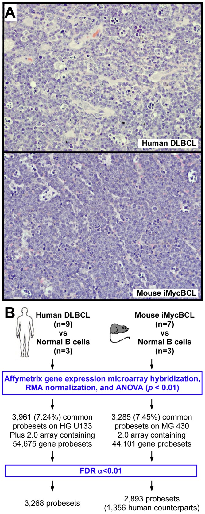
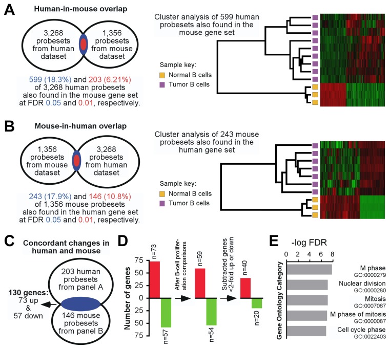
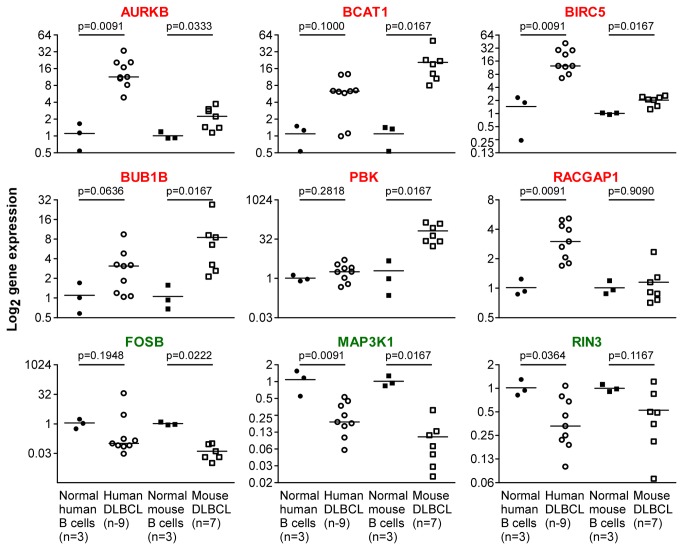
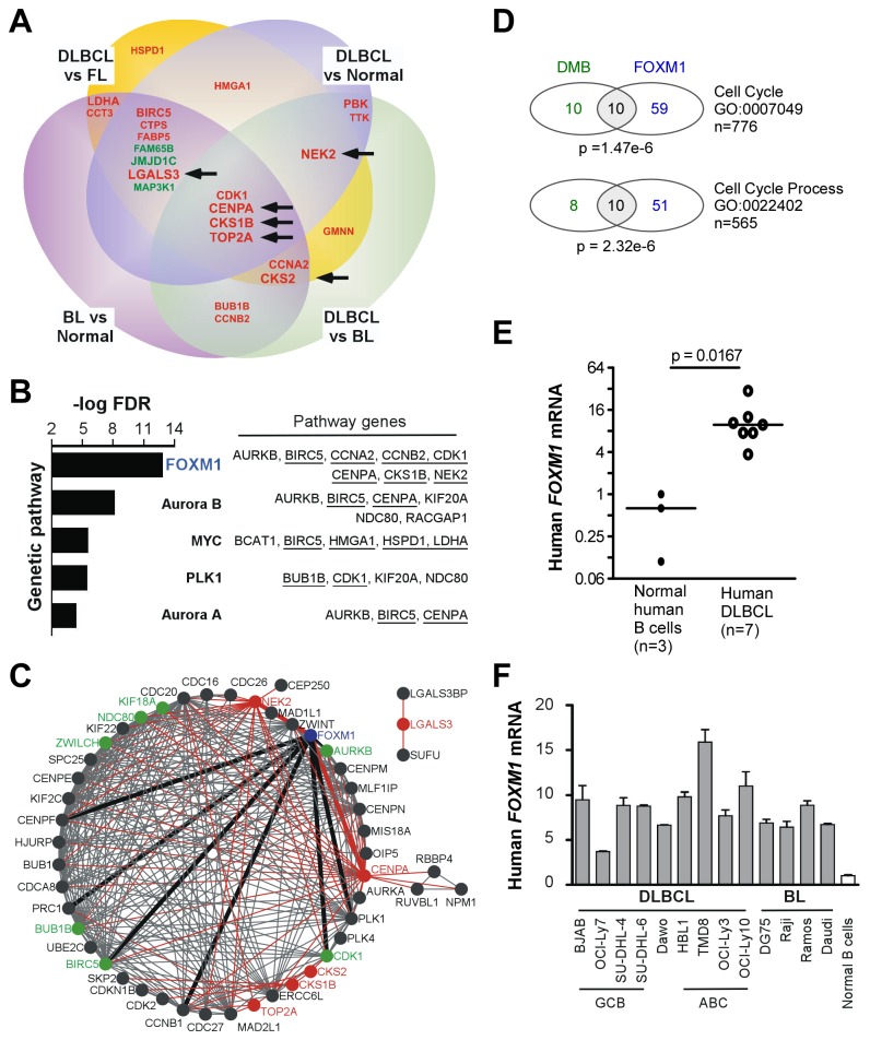
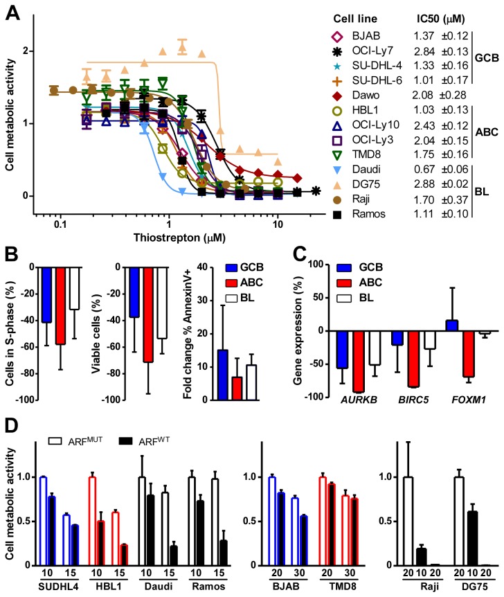
Comparative genome-wide expression profiling of malignant tumor counterparts across the human-mouse species barrier has a successful track record as a gene discovery tool in liver, breast, lung, prostate and other cancers, but has been largely neglected in studies on neoplasms of mature B-lymphocytes such as diffuse large B cell lymphoma (DLBCL) and Burkitt lymphoma (BL). We used global gene expression profiles of DLBCL-like tumors that arose spontaneously in Myc-transgenic C57BL/6 mice as a phylogenetically conserved filter for analyzing the human DLBCL transcriptome. The human and mouse lymphomas were found to have 60 concordantly deregulated genes in common, including 8 genes that Cox hazard regression analysis associated with overall survival in a published landmark dataset of DLBCL. Genetic network analysis of the 60 genes followed by biological validation studies indicate FOXM1 as a candidate DLBCL and BL gene, supporting a number of studies contending that FOXM1 is a therapeutic target in mature B cell tumors. Our findings demonstrate the value of the "mouse filter" for genomic studies of human B-lineage neoplasms for which a vast knowledge base already exists.


Conflict of interest statement

Competing Interests: The authors have declared that no competing interests exist.

Figures

Figure 1
Figure 1. Global gene expression profiles of human and mouse B lymphoma contain an abundance of deregulated genes.
(A) Representative tissue sections of human DLBCL (top) and mouse iMycBCL (bottom) stained with hematoxylin and eosin. The normal lymphoid tissue structure is effaced in both species by sheets of medium to large tumor cells that contain scant cytoplasm and round to polygonal nuclei with one to two nucleoli. Tingible body macrophages that harbor apoptotic bodies are abundant. Microscopic slides were read using an Olympus BX-51 light microscope equipped with UPLSAPO objectives (Olympus). The light temperature of the microscope bulb varied between 3000 and 3400 K. Imaging medium was air. Images were acquired with the help of a 40x objective (0.95 numerical aperture), DP2 digital camera (Olympus), and DP2-BSW imaging software (Olympus). Images were saved as TIF (tagged image file) data files and enhanced with respect to brightness, contrast and color balance using the Adobe Photoshop CS2 Version 9.0.2 software (Adobe Systems Inc). (B) Flow chart of global gene expression analysis of human and mouse lymphoma counterparts carried out in parallel and using the same statistical parameters (RMA, robust multi-averaging; ANOVA, analysis of variance; FDR, false discovery rate). Gene expression profiles of human DLBCL and normal B cells were compared on HG U133 microarrays. Gene expression profiles of mouse iMycBCL and normal B cells were compared on MG 430 microarrays.
Figure 2
Figure 2. Stringent cross-species comparison of gene expression changes in B-cell lymphoma discovers 60 concordantly deregulated genes, designated DMB (DLBCL/iMycBCL).
(A) The Venn diagram on the left shows the degree of human-in-mouse overlap of gene probesets found to be significantly variable in both species. Results at FDR threshold values of 5% and 1% are indicated in blue and red, respectively. A heat map of unsupervised cluster analysis of matching human-mouse gene sets at FDR 0.05 is depicted on the right. (B) Degree of mouse-in-human overlap of significantly variable gene probesets in both species, using the same approach as in panel A. Panels A and B depict reciprocal results. (C) Venn diagram indicating that overlapping gene sets from panels A and B (FDR 0.01 in both cases) represent 130 concordantly deregulated genes in human DLBCL and mouse iMycBCL. (D) Diagrammatic representation of two filtering steps that narrowed the gene list from 130 genes to 60 genes. The genes eliminated in this process are indicated in Table S1. (E) Column diagram indicating the top five gene ontology (GO) categories for the 130 concordantly deregulated genes from panels C and D left. GO categories were determined using DAVID. Pathway names and numbers are shown to the right.
Figure 3
Figure 3. Validation of microarray data using quantitative PCR.
Expression levels of DMB genes found to be up regulated (red; top and center rows) or down regulated (green; bottom row) on microarrays were determined with the assistance of qPCR (ΔΔCT method) in human (circle) and mouse (square) lymphoma (open) counterparts. Normal B cells (closed) were included for comparison. HPRT1 and Hprt were used as internal reference genes for human and mouse samples, respectively. Median gene expression levels are indicated by horizontal lines. Statistical analysis relied on the Mann-Whitney test.
Figure 4
Figure 4. DMB genes are part of the FOXM1 genetic network.
(A) Venn diagram displaying symbols of 23 of 60 DMB genes found to be more than 2-fold up (red) or down (green) regulated (p < 0.01) in at least 2 of 7 independent GEP studies on human lymphoma within the 4 comparison categories listed (see Table S3 for more details). Increasing font size of the gene symbol relates to an increase in the number of independent studies (2, 3, or 4) in which the gene was found to be differentially expressed across all categories. Black arrows denote the 6 genes found in 4 independent studies.(B) Column diagram indicating the result of Pathway Interaction Database (PID) analysis of the 60DMB genes. The 5 most significant pathways are shown. Genes contained in these pathways are shown to the right. Those included in panel A are underlined. (C) Network diagram representing the result of STRING analysis after expansion to a total of 50 nodes, using the 6 top genes from panel A (nodes and connecting edges indicated in red) as the only input genes. Five of 6 genes are part of a major genetic network that includes FOXM1 (node indicated in blue; edges darkened and thickened for emphasis) and 7 additional DMB genes (indicated in green). One of 6 genes, LGALS3, interacts with two other genes outside the FOXM1-associated network. (D) Venn diagrams showing the number of genes (by gene symbol) that overlap between DMB and FOXM1 target genes within the gene ontology (GO) category listed to the right. FOXM1 target genes were taken as defined by Chen et al. [37]. The given p-value indicates that FOXM1 genes are overrepresented in the DMB set and is the result of a Fisher’s Exact test to compare the proportions with a significance level set at p<0.0001. (E) qPCR result unequivocally demonstrating elevated FOXM1 mRNA in DLBCL cells (11.6 ± 3.22; open) relative to normal B cells (0.580 ± 0.258; closed). Median gene expression level is indicated by horizontal lines. Statistical analysis relied on the Mann-Whitney test. (F) Elevation of FOXM1 mRNA in DLBCL and BL cells compared to normal B cells, using qPCR as measurement tool. The classification of DLBCL lines as germinal center B cell (GCB) or activated B cell (ABC) type is indicated at the bottom. The assignment of Dawo to either of these categories is unclear.
Figure 5
Figure 5. FOXM1 inhibitors impair growth and survival of DLBCL and BL cells in vitro.
(A) Determination of the mean inhibitory concentration (IC50) of thiostrepton in 13 cell lines. Nine DLBCL and 4 BL cell lines were treated for 24 hrs with increasing concentrations of drug. Dose response curves are representative of at least three independent experiments. Cell metabolic activity was measured using the MTS assay. IC50 values and standard deviations are shown to the right. (B) Thiostrepton-dependent inhibition of cell proliferation and increased cell death. Tumor cell lines were exposed for 24 hrs to DMSO (vehicle control) or thiostrepton at IC50 levels shown in panel A, followed by flow cytometric determination of cells in S phase (DNA content analysis, left panel), number of viable cells (Guava ViaCount® analysis, middle panel), and apoptotic cells (annexin V immunoreactivity, right panel). Results are grouped by molecular subtype and shown as the average percent difference of the mean ± SD of thiostrepton- versus DMSO-treated cells. (C) Thiostrepton-dependent loss of gene expression. Cells were treated as described in panel B. RNA was isolated and gene transcript levels were measured using qPCR. Data are presented as described in panel B. (D) ARF peptide-dependent loss of cell metabolic activity. Four DLBCL and 2 BL cell lines were treated for 24 hrs with the indicated concentration (µM) of wild-type (WT) ARF peptide (black bars) or mutant (MUT) peptide (white bars) used as control. Cell metabolic activity was measured using the MTS (SUDHL4, BJAB, HBL1, TMD8) or CellTiter-Glo® (Daudi, Ramos, Raji, DG75) assays and is normalized to the mean of the lowest concentration of MUT peptide treatment per cell type. Blue, red and black outline colors indicate GCB, ABC, and BL cell lines, respectively. Data are representative of at least two independent experiments.

Similar articles

Cited by

References

    1. Lee JS, Chu IS, Mikaelyan A, Calvisi DF, Heo J et al. (2004) Application of comparative functional genomics to identify best-fit mouse models to study human cancer. Nat Genet 36: 1306-1311. doi:10.1038/ng1481. PubMed: 15565109. - DOI - PubMed
    1. Chan MM, Lu X, Merchant FM, Iglehart JD, Miron PL (2005) Gene expression profiling of NMU-induced rat mammary tumors: cross species comparison with human breast cancer. Carcinogenesis 26: 1343-1353. doi:10.1093/carcin/bgi100. PubMed: 15845649. - DOI - PubMed
    1. Sweet-Cordero A, Mukherjee S, Subramanian A, You H, Roix JJ et al. (2005) An oncogenic KRAS2 expression signature identified by cross-species gene-expression analysis. Nat Genet 37: 48-55. PubMed: 15608639. - PubMed
    1. Ellwood-Yen K, Graeber TG, Wongvipat J, Iruela-Arispe ML, Zhang J et al. (2003) Myc-driven murine prostate cancer shares molecular features with human prostate tumors. Cancer Cell 4: 223-238. doi:10.1016/S1535-6108(03)00197-1. PubMed: 14522256. - DOI - PubMed
    1. Gaspar C, Cardoso J, Franken P, Molenaar L, Morreau H et al. (2008) Cross-species comparison of human and mouse intestinal polyps reveals conserved mechanisms in adenomatous polyposis coli (APC)-driven tumorigenesis. Am J Pathol 172: 1363-1380. doi:10.2353/ajpath.2008.070851. PubMed: 18403596. - DOI - PMC - PubMed

Publication types

MeSH terms