. 2013 Oct 10;8(10):e77865.
doi: 10.1371/journal.pone.0077865. eCollection 2013.

HIV's Nef interacts with β-catenin of the Wnt signaling pathway in HEK293 cells

Affiliations

HIV's Nef interacts with β-catenin of the Wnt signaling pathway in HEK293 cells

Keren Weiser et al. PLoS One. .

Abstract

The Wnt signaling pathway is implicated in major physiologic cellular functions, such as proliferation, migration, cell fate specification, maintenance of pluripotency and induction of tumorigenicity. Proliferation and migration are important responses of T-cells, which are major cellular targets of HIV infection. Using an informatics screen, we identified a previously unsuspected interaction between HIV's Nef protein and β-catenin, a key component of the Wnt pathway. A segment in Nef contains identical amino acids at key positions and structurally mimics the β-catenin binding sites on endogenous β-catenin ligands. The interaction between Nef and β-catenin was confirmed in vitro and in a co-immunoprecipitation from HEK293 cells. Moreover, the introduction of Nef into HEK293 cells specifically inhibited a Wnt pathway reporter.


Conflict of interest statement

Competing Interests: The authors have declared that no competing interests exist.

Figures

Figure 1
Figure 1. β-catenin ligands and the identification of Nef as a novel ligand.
A. Upper panel: superimposition of known ligands of β-catenin as bound to β-catenin. β-catenin is shown in white, TCF3 is shown in green, ICAT is shown in yellow, LEF is shown in magenta, E-cadherin is shown in black, APC20 mer is shown in orange and APC15mer is shown in blue. Lower panel: same as the upper panel, however, β-catenin is not displayed. The region where the ligands converge to the same conformation is marked with a black line and titled “structural convergence” in the diagram. The two aspartates of TCF3 at the margins of the convergence are shown in x-stick representation. This region was used as the structural alignment of the ligands for the purpose of deriving the motif illustrated in panel B. B. Flow chart of the steps to identify Nef as a candidate β-catenin ligand. The chart begins with a structurally determined multiple alignment that was used to derive the β-catenin binding pattern motif. The most important residues for binding to β-catenin are colored in red. In grey, residues that were excluded from the motif as they don’t occupy the same position in 3D bound to the receptor (arginine from ICAT and lysine from APC 15 mer). Next, a Prosite-style sequence pattern was constructed to reflect the 3D structural pattern of compatibility of each residue at each specific location in the β-catenin ligand. For example, position 4 in the central region exhibits an isoleucine in some ligands and a leucine or methionine in others in contact with a hydrophobic patch on β-catenin. Thus, any of these three side chains may occupy this position and this portion of the pattern was defined as “[ILM]” to reflect this characteristic. The derived pattern then served as an input for the “MyHits” website that identified the above motif within the Nef, nucleocapsid and the MGF proteins.
Figure 2
Figure 2. Structure and sequence based evaluation of Nef.
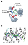
A. Superimposition of all 40 NMR structures of Nef (pdb code 2nef). The proposed β-catenin binding motif is colored blue. B. Docking of HIV1 and HIV2 Nef peptides (shown in cyan and magenta, respectively) containing the motif to β-catenin binding (shown in white). Location of the β-catenin ligand, TCF3 (shown in green), from the high-resolution crystallographic structure of the complex (pdb code: 1g3j).
Figure 3
Figure 3. Nef interacts with β-catenin.
A. Interaction of Nef and β-catenin in vitro. Purified recombinant WT-GST-Nef (or indicated D186A/F191A mutants), His- β-catenin and nickel beads mixed in physiological buffer, washed and eluted (See Methods). Top panel: immunoblot using mouse anti- β -catenin antibody (Ab) for detection. Middle panel: immunoblot using mouse anti-GST Ab for detection of Nef. Bottom panel: immunoblot using mouse anti-GST Ab for detection of Nef in total E. coli expression extract (input). This experiment was repeated three times and one representative immunoblot is shown here. B. Interaction of Nef and endogenous β-catenin in cells. HEK293 cells expressing wild-type (WT), the indicated mutants of Nef or empty vector (EV) by transfection are lysed and co-immunoprecipitated using a mouse anti- β-catenin Ab. Uppermost panel: immunoblot of immunoprecipitate (IP) using mouse anti- β-catenin Ab for detection. 2nd to top panel: immunoblot of IP using anti-TCF4 Ab for detection. 3rd to top panel: immunoblot of IP using mouse anti-Nef Ab for detection. Lowest panel: immunoblot of the total cell lysate prior to IP (input) using mouse anti-Nef Ab showing level of Nef (or mutant Nef) expression. This experiment was repeated three times and one representative immunoblot is shown here.
Figure 4
Figure 4. Nef inhibits Tcf –luciferase reporter activity.
A. Promoter negative control: HEK293 cells were transfected with the TopFlash or the FopFlash reporters and with 50 ng Nef encoding plasmid (WT-Nef, the indicated mutants or empty vector) and incubated with Wnt-conditioned media (Wnt-CM) or control media that contain no Wnt. The TopFlash plasmid has TCF binding sites that are mutated in the FopFlash plasmid. The sites in FopFlash do not interact with TCF and therefore FopFlash serves as a negative control. Renilla luciferase was used for normalization purposes. Data shown here represent two experiments, each done in triplicate. WT-Nef significantly (P<0.00001, t-test) inhibits TCF reporter activity as compared to empty vector and also compared to the β-catenin motif mutants, D186A-Nef and F191A-Nef (P<0.00001, t-test) when co-transfected with TopFlash. B. The firefly/renilla values from panel A of this figure were used to calculate the TopFlash/FopFlash ratios of each condition. The resultant TopFlash/FopFlash ratios were then used to calculate the percent inhibition by WT-Nef and the indicated mutants on transcription in cells stimulated with Wnt.
Figure 5
Figure 5. Model of Nef in situ on β-catenin.
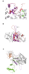
A. The C-terminal tail of Nef after position 185 is rearranged from its unbound position into the conformation seen in β-catenin ligands. The curved arrow shows the hypothesized trajectory of the rearrangement. The backbone of un-rearranged Nef is shown in red ribbon, re-arranged Nef in magenta, β-catenin in grey, bound E-cadherin in yellow. The key hotspots (Asp674/ Phe679 of E-cadherin and Asp186/Phe191 of Nef) are displayed in stick depiction. B. A different view of the complex described in 5A. The black arrow points to the loop in Nef that is important for binding to adaptor proteins, MHC proteins, and is also important in sorting these proteins into clathrin-coated pits ([10,15,85,86]). C. The β-catenin bound form of Nef (magenta) is superimposed with a dimeric Nef conformation (green) bound to the Fyn SH3 domain (orange; pdb 1avz, [87]).

Similar articles

Cited by

References

    1. Kestler HW 3rd, Ringler DJ, Mori K, Panicali DL, Sehgal PK et al. (1991) Importance of the nef gene for maintenance of high virus loads and for development of AIDS. Cell 65: 651-662. doi:10.1016/0092-8674(91)90097-I. PubMed: 2032289. - DOI - PubMed
    1. de Ronde A, Klaver B, Keulen W, Smit L, Goudsmit J (1992) Natural HIV-1 NEF accelerates virus replication in primary human lymphocytes. Virology 188: 391-395. doi:10.1016/0042-6822(92)90772-H. PubMed: 1566581. - DOI - PubMed
    1. Kirchhoff F, Münch J, Carl S, Stolte N, Mätz-Rensing K et al. (1999) The human immunodeficiency virus type 1 nef gene can to a large extent replace simian immunodeficiency virus nef in vivo. J Virol 73: 8371-8383. PubMed: 10482588. - PMC - PubMed
    1. Deacon NJ, Tsykin A, Solomon A, Smith K, Ludford-Menting M et al. (1995) Genomic structure of an attenuated quasi species of HIV-1 from a blood transfusion donor and recipients. Science 270: 988-991. doi:10.1126/science.270.5238.988. PubMed: 7481804. - DOI - PubMed
    1. Churchill MJ, Rhodes DI, Learmont JC, Sullivan JS, Wesselingh SL et al. (2006) Longitudinal analysis of human immunodeficiency virus type 1 nef/long terminal repeat sequences in a cohort of long-term survivors infected from a single source. J Virol 80: 1047-1052. doi:10.1128/JVI.80.2.1047-1052.2006. PubMed: 16379007. - DOI - PMC - PubMed

Publication types

Substances