Divergent modification of low-dose ⁵⁶Fe-particle and proton radiation on skeletal muscle
- PMID: 24131063
- PMCID: PMC4152935
- DOI: 10.1667/RR3329.1
Divergent modification of low-dose ⁵⁶Fe-particle and proton radiation on skeletal muscle
Abstract
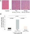
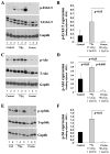
It is unknown whether loss of skeletal muscle mass and function experienced by astronauts during space flight could be augmented by ionizing radiation (IR), such as low-dose high-charge and energy (HZE) particles or low-dose high-energy proton radiation. In the current study adult mice were irradiated whole-body with either a single dose of 15 cGy of 1 GeV/n ⁵⁶Fe-particle or with a 90 cGy proton of 1 GeV/n proton particles. Both ionizing radiation types caused alterations in the skeletal muscle cytoplasmic Ca²⁺ ([Ca²⁺]i) homeostasis. ⁵⁶Fe-particle irradiation also caused a reduction of depolarization-evoked Ca²⁺ release from the sarcoplasmic reticulum (SR). The increase in the [Ca²⁺]i was detected as early as 24 h after ⁵⁶Fe-particle irradiation, while effects of proton irradiation were only evident at 72 h. In both instances [Ca²⁺]i returned to baseline at day 7 after irradiation. All ⁵⁶Fe-particle irradiated samples revealed a significant number of centrally localized nuclei, a histologic manifestation of regenerating muscle, 7 days after irradiation. Neither unirradiated control or proton-irradiated samples exhibited such a phenotype. Protein analysis revealed significant increase in the phosphorylation of Akt, Erk1/2 and rpS6k on day 7 in ⁵⁶Fe-particle irradiated skeletal muscle, but not proton or unirradiated skeletal muscle, suggesting activation of pro-survival signaling. Our findings suggest that a single low-dose ⁵⁶Fe-particle or proton exposure is sufficient to affect Ca²⁺ homeostasis in skeletal muscle. However, only ⁵⁶Fe-particle irradiation led to the appearance of central nuclei and activation of pro-survival pathways, suggesting an ongoing muscle damage/recovery process.
Figures


Similar articles
-
Cardiovascular risks associated with low dose ionizing particle radiation.PLoS One. 2014 Oct 22;9(10):e110269. doi: 10.1371/journal.pone.0110269. eCollection 2014. PLoS One. 2014. PMID: 25337914 Free PMC article.
-
Executive function in rats is impaired by low (20 cGy) doses of 1 GeV/u (56)Fe particles.Radiat Res. 2012 Oct;178(4):289-94. doi: 10.1667/rr2862.1. Epub 2012 Aug 10. Radiat Res. 2012. PMID: 22880624
-
Different Sequences of Fractionated Low-Dose Proton and Single Iron-Radiation-Induced Divergent Biological Responses in the Heart.Radiat Res. 2017 Aug;188(2):191-203. doi: 10.1667/RR14667.1. Epub 2017 Jun 14. Radiat Res. 2017. PMID: 28613990 Free PMC article.
-
Fundamental space radiobiology.Gravit Space Biol Bull. 2003 Jun;16(2):29-36. Gravit Space Biol Bull. 2003. PMID: 12959129 Review.
-
Space radiation health: a brief primer.Gravit Space Biol Bull. 2003 Jun;16(2):1-4. Gravit Space Biol Bull. 2003. PMID: 12959125 Review.
Cited by
-
Effect of irradiation on Akt signaling in atrophying skeletal muscle.J Appl Physiol (1985). 2016 Oct 1;121(4):917-924. doi: 10.1152/japplphysiol.00218.2016. Epub 2016 Aug 25. J Appl Physiol (1985). 2016. PMID: 27562841 Free PMC article.
-
Influence of longitudinal radiation exposure from microcomputed tomography scanning on skeletal muscle function and metabolic activity in female CD-1 mice.Physiol Rep. 2017 Jul;5(13):e13338. doi: 10.14814/phy2.13338. Physiol Rep. 2017. PMID: 28676556 Free PMC article.
-
Long-Term Effects of Very Low Dose Particle Radiation on Gene Expression in the Heart: Degenerative Disease Risks.Cells. 2021 Feb 13;10(2):387. doi: 10.3390/cells10020387. Cells. 2021. PMID: 33668521 Free PMC article.
References
-
- Grigoryeva LS, Kozlovskaya IB. Effect of weightlessness and hypokinesis on velocity and strength properties of human muscles. Kosmicheskaya Biologiya I Aviakosmicheskaya Meditsina. 1987;21:27–30. - PubMed
-
- Edgerton VR, Zhou MY, Ohira Y, Klitgaard H, Jiang B, Bell G, et al. Human fiber size and enzymatic properties after 5 and 11 days of spaceflight. J Appl Physiol. 1995;78:1733–9. - PubMed
-
- Stevens L, Mounier Y. Ca2+ movements in sarcoplasmic reticulum of rat soleus fibers after hindlimb suspension. J Appl Physiol. 1992;72:1735–40. - PubMed
-
- Caiozzo VJ, Haddad F, Baker MJ, Herrick RE, Prietto N, Baldwin KM. Microgravity-induced transformations of myosin isoforms and contractile properties of skeletal muscle. J Appl Physiol. 1996;81:123–32. - PubMed
-
- Roy RR, Hodgson JA, Aragon J, Day MK, Kozlovskaya I, Edgerton VR. Recruitment of the Rhesus soleus and medial gastrocnemius before, during and after spaceflight. J Gravit Physiol. 1996;3:11–5. - PubMed
Publication types
MeSH terms
Substances
Grants and funding
LinkOut - more resources
Full Text Sources
Other Literature Sources
Research Materials
Miscellaneous

