

Lysophosphatidic acid protects human mesenchymal stromal cells from differentiation-dependent vulnerability to apoptosis
- PMID: 24131310
- PMCID: PMC3993074
- DOI: 10.1089/ten.TEA.2013.0487
Lysophosphatidic acid protects human mesenchymal stromal cells from differentiation-dependent vulnerability to apoptosis
Abstract
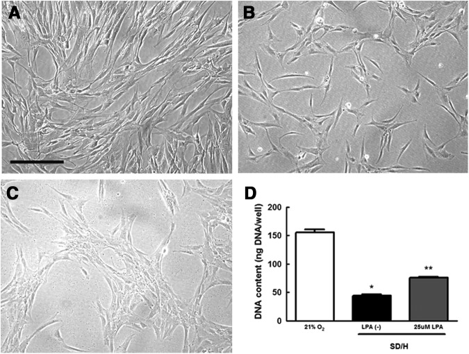
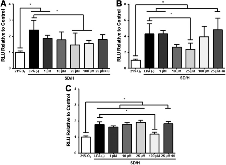
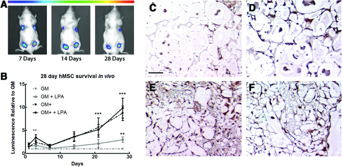
The survival of transplanted cells and their resulting efficacy in cell-based therapies is markedly impaired due to serum deprivation and hypoxia (SD/H) resulting from poor vascularization within tissue defects. Lysophosphatidic acid (LPA) is a platelet-derived growth factor with pleiotropic effects on many cell types. Mesenchymal stromal cells (MSC) exhibit unique secretory and stimulatory characteristics depending on their differentiation state. In light of the potential of MSC in cell-based therapies, we examined the ability of LPA to abrogate SD/H-induced apoptosis in human MSC at increasing stages of osteogenic differentiation in vitro and assessed MSC survival in vivo. Undifferentiated MSC were rescued from SD/H-induced apoptosis by treatment with both 25 and 100 μM LPA. However, MSC conditioned with osteogenic supplements responded to 25 μM LPA, and cells conditioned with dexamethasone-containing osteogenic media required 100 μM LPA. This rescue was mediated through LPA1 in all cases. The addition of 25 μM LPA enhanced vascular endothelial growth factor (VEGF) secretion by MSC in all conditions, but VEGF availability was not responsible for protection against apoptosis. We also showed that codelivery of 25 μM LPA with MSC in alginate hydrogels significantly improved the persistence of undifferentiated MSC in vivo over 4 weeks as measured by bioluminescence imaging. Osteogenic differentiation alone was protective of SD/H-induced apoptosis in vitro, and the synergistic delivery of LPA did not enhance persistence of osteogenically induced MSC in vivo. These data demonstrate that the capacity of LPA to inhibit SD/H-induced apoptosis in MSC is dependent on both the differentiation state and dosage. This information will be valuable for optimizing osteogenic conditioning regimens for MSC before in vivo implementation.
Figures

Similar articles
-
Lysophosphatidic acid protects mesenchymal stem cells against ischemia-induced apoptosis in vivo.Stem Cells Dev. 2009 Sep;18(7):947-54. doi: 10.1089/scd.2008.0352. Stem Cells Dev. 2009. PMID: 19193014
-
LPA rescues ER stress-associated apoptosis in hypoxia and serum deprivation-stimulated mesenchymal stem cells.J Cell Biochem. 2010 Nov 1;111(4):811-20. doi: 10.1002/jcb.22731. J Cell Biochem. 2010. PMID: 20533299
-
Lysophosphatidic acid enhances human umbilical cord mesenchymal stem cell viability without differentiation via LPA receptor mediating manner.Apoptosis. 2017 Oct;22(10):1296-1309. doi: 10.1007/s10495-017-1399-6. Apoptosis. 2017. PMID: 28766061 Free PMC article.
-
Lysophosphatidic acid: Its role in bone cell biology and potential for use in bone regeneration.Prostaglandins Other Lipid Mediat. 2019 Aug;143:106335. doi: 10.1016/j.prostaglandins.2019.106335. Epub 2019 May 1. Prostaglandins Other Lipid Mediat. 2019. PMID: 31054330 Review.
-
Revisiting the role of lysophosphatidic acid in stem cell biology.Exp Biol Med (Maywood). 2021 Aug;246(16):1802-1809. doi: 10.1177/15353702211019283. Epub 2021 May 26. Exp Biol Med (Maywood). 2021. PMID: 34038224 Free PMC article. Review.
Cited by
-
Role of TAZ in Lysophosphatidic Acid-Induced Migration and Proliferation of Human Adipose-Derived Mesenchymal Stem Cells.Biomol Ther (Seoul). 2017 Jul 1;25(4):354-361. doi: 10.4062/biomolther.2016.263. Biomol Ther (Seoul). 2017. PMID: 28554198 Free PMC article.
-
Cellularizing hydrogel-based scaffolds to repair bone tissue: How to create a physiologically relevant micro-environment?J Tissue Eng. 2017 Jun 8;8:2041731417712073. doi: 10.1177/2041731417712073. eCollection 2017 Jan-Dec. J Tissue Eng. 2017. PMID: 28634532 Free PMC article.
-
Detection of Pentosidine Cross-Links in Cell-Secreted Decellularized Matrices Using Time Resolved Fluorescence Spectroscopy.ACS Biomater Sci Eng. 2017 Sep 11;3(9):1944-1954. doi: 10.1021/acsbiomaterials.6b00029. Epub 2016 May 31. ACS Biomater Sci Eng. 2017. PMID: 28944287 Free PMC article.
-
Lysophosphatidic acid enhances survival of human CD34(+) cells in ischemic conditions.Sci Rep. 2015 Nov 10;5:16406. doi: 10.1038/srep16406. Sci Rep. 2015. PMID: 26553339 Free PMC article.
-
Human mesenchymal stem cell spheroids in fibrin hydrogels exhibit improved cell survival and potential for bone healing.Cell Tissue Res. 2014 Jul;357(1):91-9. doi: 10.1007/s00441-014-1830-z. Epub 2014 Apr 30. Cell Tissue Res. 2014. PMID: 24781147 Free PMC article.
References
-
- Caplan A.I.Adult mesenchymal stem cells for tissue engineering versus regenerative medicine. J Cell Physiol 213,341, 2007 - PubMed
-
- Peter S.J., Liang C.R., Kim D.J., Widmer M.S., and Mikos A.G.Osteoblastic phenotype of rat marrow stromal cells cultured in the presence of dexamethasone, beta-glycerolphosphate, and L-ascorbic acid. J Cell Biochem 71,55, 1998 - PubMed
Publication types
MeSH terms
Substances
Grants and funding
LinkOut - more resources
Full Text Sources
Other Literature Sources
Miscellaneous
