. 2013 Oct 16:14:123.
doi: 10.1186/1471-2202-14-123.

Structural brain abnormalities in cervical dystonia

Affiliations

Structural brain abnormalities in cervical dystonia

Tino Prell et al. BMC Neurosci. .

Abstract

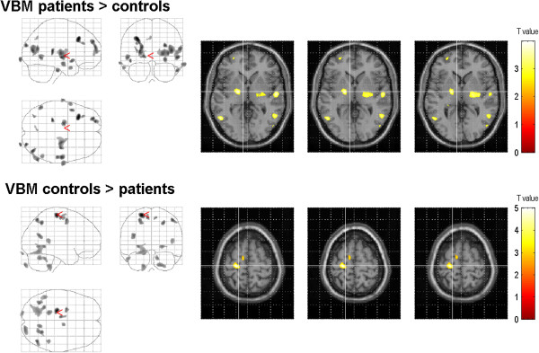
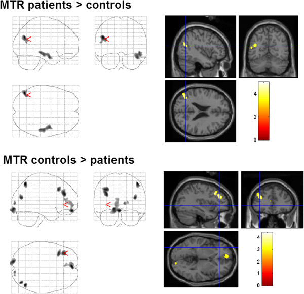
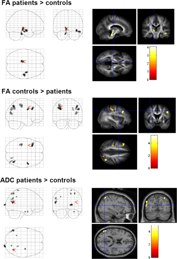
Background: Idiopathic cervical dystonia is characterized by involuntary spasms, tremors or jerks. It is not restricted to a disturbance in the basal ganglia system because non-conventional voxel-based MRI morphometry (VBM) and diffusion tensor imaging (DTI) have detected numerous regional changes in the brains of patients.In this study scans of 24 patients with cervical dystonia and 24 age-and sex-matched controls were analysed using VBM, DTI and magnetization transfer imaging (MTI) using a voxel-based approach and a region-of-interest analysis. Results were correlated with UDRS, TWSTRS and disease duration.

Results: We found structural alterations in the basal ganglia; thalamus; motor cortex; premotor cortex; frontal, temporal and parietal cortices; visual system; cerebellum and brainstem of the patients with dystonia.

Conclusions: Cervical dystonia is a multisystem disease involving several networks such as the motor, sensory and visual systems.


Figures

Figure 1
Figure 1
Changes of grey matter volume in patients with dystonia compared to those in healthy controls (group comparison). VBM showed regional enhanced grey matter in the lentiform nucleus, the frontal eye fields (BA 8), and the secondary visual cortex (displayed at p = 0.001, uncorrected, extent threshold 100 voxels). Grey matter atrophy was observed in the precentral gyrus, the supplementary motor area, the somatosensory association cortex, and in the medial temporal gyrus. The color bar represents the T-score. The differences between the groups are superimposed on a standard normalized T1-weighted image. Images are shown in neurological convention.
Figure 2
Figure 2
Changes of MTR in patients with dystonia compared to those in healthy controls (group comparison). Enhanced MTR was found in the left temporal medial gyrus, in the temporal lobe, in the secondary somatosensory cortex, and in the cingulate gyrus. MTR was reduced in the left dorsolateral prefrontal and frontal cortex (BA 9, 10, 11) and in the primary and secondary visual cortex (BA 17, 19).
Figure 3
Figure 3
Changes of DTI-values in patients with dystonia compared to those in healthy controls (group comparison). FA was increased in brainstem, thalamus, and temporal areas. Decreased FA was observed in postcentral gyrus, frontal and occipital cortex. Diffusivity was enhanced in the area MT, and secondary somatosensory cortex.

Similar articles

Cited by

References

    1. Defazio G, Abbruzzese G, Livrea P, Berardelli A. Epidemiology of primary dystonia. Lancet Neurol. 2004;3:673–678. doi: 10.1016/S1474-4422(04)00907-X. - DOI - PubMed
    1. Chan J, Brin MF, Fahn S. Idiopathic cervical dystonia: clinical characteristics. Mov Disord. 1991;6:119–126. doi: 10.1002/mds.870060206. - DOI - PubMed
    1. Draganski B, Thun-Hohenstein C, Bogdahn U, Winkler J, May A. “Motor circuit” gray matter changes in idiopathic cervical dystonia. Neurology. 2003;61:1228–1231. doi: 10.1212/01.WNL.0000094240.93745.83. - DOI - PubMed
    1. Obermann M, Yaldizli O, De Greiff A, Lachenmayer ML, Buhl AR, Tumczak F, Gizewski ER, Diener HC, Maschke M. Morphometric changes of sensorimotor structures in focal dystonia. Mov Disord. 2007;22:1117–1123. doi: 10.1002/mds.21495. - DOI - PubMed
    1. Egger K, Mueller J, Schocke M, Brenneis C, Rinnerthaler M, Seppi K, Trieb T, Wenning GK, Hallett M, Poewe W. Voxel based morphometry reveals specific gray matter changes in primary dystonia. Mov Disord. 2007;22:1538–1542. doi: 10.1002/mds.21619. - DOI - PubMed

Publication types

LinkOut - more resources