. 2013 Nov;140(22):4522-32.
doi: 10.1242/dev.092775. Epub 2013 Oct 16.

Fetal adrenal capsular cells serve as progenitor cells for steroidogenic and stromal adrenocortical cell lineages in M. musculus

Affiliations

Fetal adrenal capsular cells serve as progenitor cells for steroidogenic and stromal adrenocortical cell lineages in M. musculus

Michelle A Wood et al. Development. 2013 Nov.

Abstract

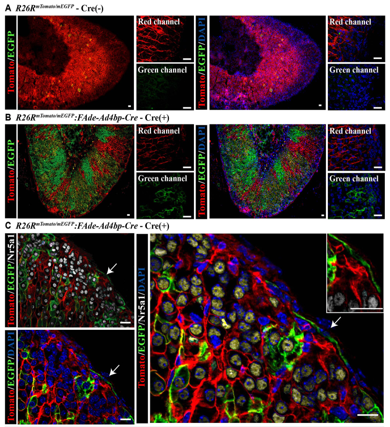
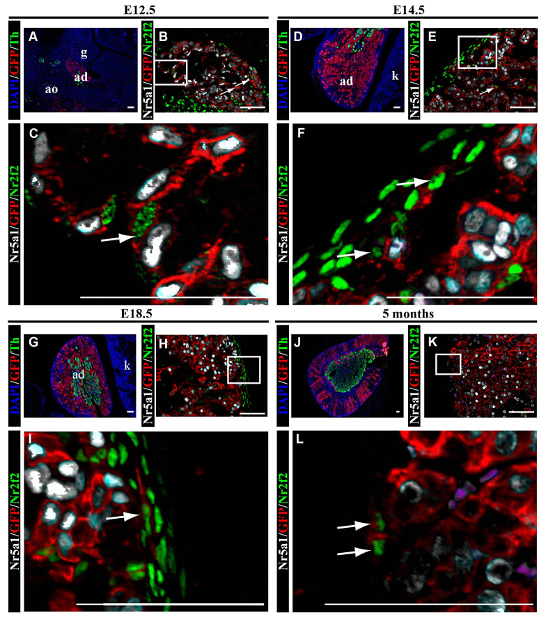
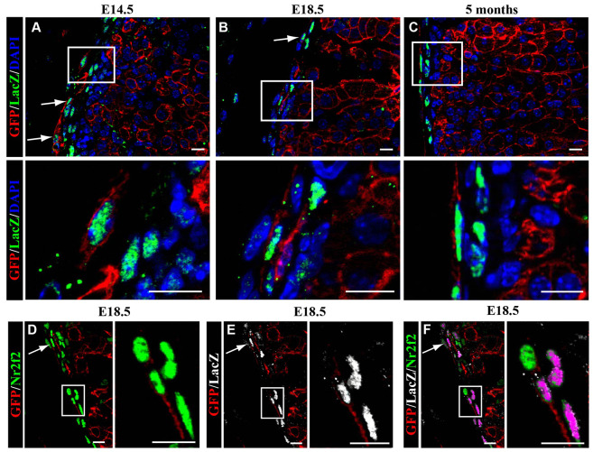
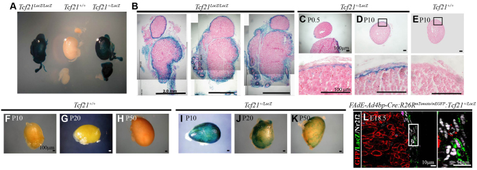
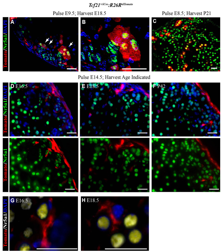
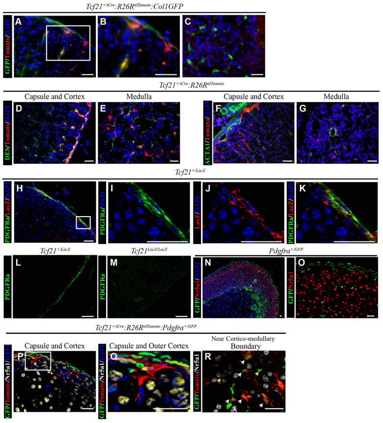
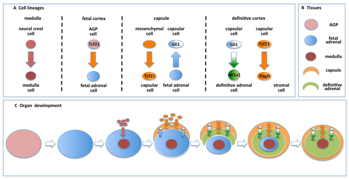
The lineage relationships of fetal adrenal cells and adrenal capsular cells to the differentiated adrenal cortex are not fully understood. Existing data support a role for each cell type as a progenitor for cells of the adult cortex. This report reveals that subsets of capsular cells are descendants of fetal adrenocortical cells that once expressed Nr5a1. These fetal adrenocortical cell descendants within the adrenal capsule express Gli1, a known marker of progenitors of steroidogenic adrenal cells. The capsule is also populated by cells that express Tcf21, a known inhibitor of Nr5a1 gene expression. We demonstrate that Tcf21-expressing cells give rise to Nr5a1-expressing cells but only before capsular formation. After the capsule has formed, capsular Tcf21-expressing cells give rise only to non-steroidogenic stromal adrenocortical cells, which also express collagen 1a1, desmin and platelet-derived growth factor (alpha polypeptide) but not Nr5a1. These observations integrate prior observations that define two separate origins of adult adrenocortical steroidogenic cells (fetal adrenal cortex and/or the adrenal capsule). Thus, these observations predict a unique temporal and/or spatial role of adult cortical cells that arise directly from either fetal cortical cells or from fetal cortex-derived capsular cells. Last, the data uncover the mechanism by which two populations of fetal cells (fetal cortex derived Gli1-expressing cells and mesenchymal Tcf21-expressing mesenchymal cells) participate in the establishment of the homeostatic capsular progenitor cell niche of the adult cortex.

Keywords: Adrenal gland; Homeostasis; Mouse; Progenitor cells.


Figures

Fig. 1.
Fig. 1.
Fetal adrenal cells give rise to capsular and steroidogenic cells of the adult adrenal gland. Using immunofluorescence (IF), cryosections from adult FAdE-Ad4bp-Cre:R26RmTomato/mEGFP mice reveal that in the absence of Cre (A), the ubiquitous Tomato reporter (red membrane R26RmTomato/mEGFP without Cre activation) is expressed throughout the gland but in Cre-expressing littermates (B), EGFP reporter expression (green membrane R26RmTomato/mEGFP:FAdE-Ad4bp-Cre) is detected. Largest panels show red (549 nm excitation wavelength) and green (488 nm excitation wavelength) channels overlaid. High-power magnification insets on the right show each channel individually. (C) EGFP-expressing cells give rise to both Nr5a1-expressing cells in the cortex (white nuclei) and Nr5a1-negative cells in the adrenal capsule (arrows and inset). Scale bars: 20 μm.
Fig. 2.
Fig. 2.
Fetal adrenal cells give rise to cells of the adrenal capsule. Adrenal glands from FAdE-Ad4bp-Cre:R26RmTomato/mEGFP mice were evaluated by immunofluorescence in paraffin sections. In the fetal adrenal gland, at E12.5 (A), medullary cells expressing Th (green cytoplasm) and adrenocortical cells expressing EGFP (red membrane; R26RmTomato/mEGFP:FAdE-Ad4bp-Cre) can be seen intermingling. EGFP-positive cells are indicative of fetal adrenocortical cell lineage (FAdE-Ad4bp-Cre expressing). (B) Low power and (C) high-power magnifications showing that Nr2f2-expressing capsular cells (green nuclei) and Nr5a1-expressing fetal adrenocortical cells (white nuclei) are intermingled. Both cell types co-express EGFP (red membrane, arrows), indicative of fetal adrenocortical lineage. (D) By E14.5, adrenal capsule formation and neural crest cell migration to the medulla, as reflected by Th expression (green cytoplasm) is complete. (E) Low-power and (F) high-power magnifications showing Nr2f2-expressing cells (green nuclei) are primarily in the adrenal capsule and some of these cells contain EGFP (red membrane), which is indicative of fetal adrenocortical lineage. These capsular cells expressing both Nr2f2 (green nuclei) and EGFP (red membrane) can be found in animals at all ages examined (white arrows) - E18.5 (G-I) and 5 months (J-L). (L) Magenta cells are red blood cells that are autofluorescent in every channel but do not stain with DAPI. C,F,I and L are enlargements of boxes in B,E,H and K, respectively. Blue in A,D,G and J indicates DAPI. Scale bars: 50 μm. Cyan results from overlay of white and red. ad, adrenal gland; g, gonad; ao, aorta; k, kidney.
Fig. 3.
Fig. 3.
Capsular descendants of the fetal adrenal cortex express Gli1. Adrenal glands from FAdE-Ad4bp-Cre:R26RmTomato/mEGFP:Gli1-LacZ mice were evaluated by immunofluorescence in paraffin sections. At all ages examined, E14.5 (A), E18.5 (B,D,F) and at 5 months (C), some cells expressing β-gal (LacZ, green nuclei; indicative of Gli1 promoter activity) also express EGFP (red membrane; indicative of fetal adrenocortical lineage, R26RmTomato/mEGFP:FAdE-Ad4bp-Cre expressing). Bottom panels in A-C are enlargements of boxed areas in the top panels. White arrows indicate cells expressing both β-gal (Gli1-LacZ, green nuclei) and EGFP (red membrane). (D-F) In the capsule of an E18.5 adrenal gland, EGFP-expressing cells (red membrane) co-express both Nr2f2 (green nuclei; D) and β-gal (Gli1-LacZ, white nuclei; E) with triple overlay in F. Magenta indicates overlap of green (Nr2f2) and white (Gli1-LacZ). Boxed areas in D-F are shown enlarged in the right panels. White arrows in D and E indicate cells expressing both markers. In F, white arrow indicates cells expressing EGFP, LacZ and Nr2f2. Scale bars: 10 μm.
Fig. 4.
Fig. 4.
Tcf21 is expressed in the adrenal capsule but does not colocalize with descendants of fetal adrenocortical cells. Adrenal glands harvested from Tcf21+/+, Tcf21+/LacZ or Tcf21LacZ/LacZ mice were evaluated by whole-mount β-gal staining to characterize Tcf21 promoter activity during adrenal gland development. (A) Whole-mount staining of adrenal gland, kidney and gonad from male littermates of each genotype at P0.5 show β-gal activity in +/LacZ and LacZ/LacZ animals. (B) Improper separation of the adrenal gland and gonad can be seen in organs from Tcf21LacZ/LacZ mice. All panels are from the same adrenal, gonad and kidney unit. Scale bars: 2 mm. Cross-sections of P0.5 (C) and P10 (D) adrenal glands reveal staining predominantly in the capsule with a few positive cells in the adrenal cortex when compared with wild-type controls (Tcf21+/+; E). Postnatal Tcf21+/+ (wild type) adrenal glands lack β-gal activity at P10 (F), P20 (G) and P50 (H). Postnatal Tcf21+/LacZ adrenal glands show decreasing capsular β-gal activity from P10 (I) through P20 (J) and P50 (K). All cross-sections were counterstained with Eosin (pink). Scale bars: 100 μm. (L) Adrenal glands from FAdE-Ad4bp-Cre:R26RmTomato/mEGFP:Tcf21+/LacZ mice were used for lineage tracing and evaluation by immunofluorescence in paraffin sections. EGFP expression (red membrane; R26RmTomato/mEGFP:FAdE-Ad4bp-Cre) in descendants of fetal adrenal cells (FAdE-Ad4bp-Cre expressing) did not overlap with β-gal expression (LacZ, green cytoplasm; Tcf21-expressing cells; Tcf21+/LacZ), despite both EGFP and β-gal co-localizing individually with Nr2f2 (white nuclei). Panel on the right is an enlargement of the box in the left panel. Scale bars: 10 μm. Abbreviations: ad, adrenal gland; g, gonad; k, kidney.
Fig. 5.
Fig. 5.
Tcf21-expressing cells give rise to steroidogenic adrenocortical cells prior to adrenal capsule formation and non-steroidogenic adrenocortical cells after adrenal capsule formation. Adrenal glands from Tcf21+/iCre:R26RtdTomato mice were harvested at E18.5 after tamoxifen induction at E9.5 (prior to adrenal capsule formation) and were evaluated by immunofluorescence in cryosections (A,B). Tomato-expressing (red cytoplasm; Tcf21+/iCre:R26RtdTomato) descendants of Tcf21-expressing cells were found both in the capsule and in a few adrenocortical cells that expressed Nr5a1 (green nucleus, white arrows). (B) An enlargement of A. Similar to A and B, in C the adrenal glands were harvested at P21 after tamoxifen induction at E8.5. (A-C) Yellow indicates overlap of red and green. Arrows in A indicate cells expressing both Nr5a1 and tomato, and thus descendants of Tcf21-expressing cells. (D-H) Adrenal glands from Tcf21+/iCre:R26RtdTomato mice harvested at various times after administration of tamoxifen to pregnant females at E14.5 (after adrenal capsule formation). Adrenocortical cells failed to show co-expression of Nr5a1 (green nuclei) and Tomato (red cytoplasm) at E16.5 (D), E18.5 (E) and P42 (F). In G (E16.5) and H (E18.5), Tomato expression (red cytoplasm) belongs to cells lacking Nr5a1 expression (white nuclei). (G,H) Yellow tint indicates overlap of white and blue staining. DAPI was used to visualize nuclei in all panels (blue). Scale bars: 20 μm.
Fig. 6.
Fig. 6.
Tcf21-expressing cells give rise to stromal cells after adrenal capsule formation. (A-C) Adrenal glands from 8-week-old Tcf21+/iCre:R26RtdTomato:ColGFP mice were harvested after a 2-week induction with tamoxifen starting at 4 weeks of age. Adrenal glands analyzed by immunofluorescence of cryosections. GFP (green, stromal cells; Col1GFP) and Tomato (red, Tcf21 lineage; Tcf21+/iCre:R26RtdTomato) were found to colocalize (yellow) in both the adrenal capsule and in spindle-shaped cells of the cortex (A,B) and medulla (C). (B) An enlargement of the boxed area in A. (D-G) Adrenal glands from Tcf21+/iCre:R26RtdTomato were harvested at P21 after a single tamoxifen induction at P7 and analyzed by immunofluorescence in cryosections. Desmin (DES, green) and Tomato (red, Tcf21-lineage) were found to colocalize in the adrenal capsule, cortex (D) and medulla (E). Smooth muscle actin (ACTA, green) and Tomato (red, Tcf21-lineage) were found in close proximity but do not appear to colocalize (F,G). (H-M) Adrenal glands from P0.5 Tcf21+/LacZ mice were harvested, embedded in paraffin and analyzed by immunofluorescence to characterize Pdgfra expression. Pdgfra (green membrane) is predominantly present in the adrenal capsule with Tcf21 (as detected by anti-β-gal antibody; red cytoplasm; H,I; Tcf21+/LacZ). Both Pdgfra (I, green membrane) and β-gal (J, LacZ, red cytoplasm) appear to be co-expressed in the adrenal capsule (K). (I-K) Enlargements of boxed area in H. Pdgrfa expression (green) is diminished in adrenal glands from Tcf21LacZ/LacZ mice (M) when compared with wild type (L). (N,O) Immunofluorescence on paraffin sections of adrenal glands from Pdgfrα+/GFP reporter mice reveal GFP protein (green nuclei; Pdgfrα+/GFP) in the adrenal capsule and some cells of the adrenal cortex but not in steroidogenic Nr5a1-expressing cells (red nuclei). Large cytomegalic cells at the corticomedullary boundary are common in adult adrenal glands and are autofluorescent. (P-R) Adrenal glands were harvested from Tcf21+/iCre:R26RtdTomato:Pdgfrα+/GFP mice at E18.5 after tamoxifen administration at E14.5 and analyzed by immunofluorescence in cryosections. Tomato expression (indicative of Tcf21 lineage; red cytoplasm) was colocalized with GFP (green nuclei) in Pdgfra-expressing cells of the adrenal capsule and cortex but did not colocalize with Nr5a1-expressing adrenocortical cells (white nuclei). (Q) Enlargement of the boxed area in P. Tomato- and GFP-expressing cells could be found throughout the cortex, including close to the corticomedullary boundary (R). White arrows indicate cells co-expressing Tomato and GFP; yellow arrows indicate GFP-expressing cells without Tomato. Nuclei in all panels except L, M and O are visualized with DAPI. Scale bars: 20 μm.
Fig. 7.
Fig. 7.
Model of adrenal lineage and homeostasis. (A) Multiple cell lineages contribute to cells of the differentiated adrenal gland. Neural crest cells (red) give rise to catecholamine-secreting cells of the adrenal medulla. Tcf21-expressing cells contribute to the pool of fetal adrenal cells and arise from a mesenchymal cell lineage prior to contributing to the adrenal capsule (orange). Fetal adrenal cells (blue) contribute to a subpopulation of capsular cells that express Gli1 (white). Gli1-expressing capsular cells give rise to steroidogenic adrenocortical cells (green) of the adult gland, whereas Tcf21-expressing capsular cells give rise to Pdgfr-expressing stromal adrenocortical cells (orange). (B) The four tissue lineages represented in C. (C) During adrenal organogenesis starting with adrenogonadal primordia (AGP), cells from the neural crest invade the fetal adrenal primordia to form the medulla, and mesenchymal cells contribute to the adrenal capsule. Fetal adrenal cells contribute to the adrenal capsule. As the adult cortex replaces the fetal cortex, Tcf21-expressing and Gli1-expressing cells serve as progenitors in the adrenal capsule that give rise to stromal and steroidogenic adrenocortical cells, respectively. Upon completion of organogenesis, the capsular progenitor cells are retained throughout adulthood and contribute to homeostatic maintenance of the adrenal cortex.

References

    1. Acharya A., Baek S. T., Banfi S., Eskiocak B., Tallquist M. D. (2011). Efficient inducible Cre-mediated recombination in Tcf21 cell lineages in the heart and kidney. Genesis 49, 870–877 - PMC - PubMed
    1. Acharya A., Baek S. T., Huang G., Eskiocak B., Goetsch S., Sung C. Y., Banfi S., Sauer M. F., Olsen G. S., Duffield J. S., et al. (2012). The bHLH transcription factor Tcf21 is required for lineage-specific EMT of cardiac fibroblast progenitors. Development 139, 2139–2149 - PMC - PubMed
    1. Ahn S., Joyner A. L. (2004). Dynamic changes in the response of cells to positive hedgehog signaling during mouse limb patterning. Cell 118, 505–516 - PubMed
    1. Bai C. B., Auerbach W., Lee J. S., Stephen D., Joyner A. L. (2002). Gli2, but not Gli1, is required for initial Shh signaling and ectopic activation of the Shh pathway. Development 129, 4753–4761 - PubMed
    1. Bingham N. C., Verma-Kurvari S., Parada L. F., Parker K. L. (2006). Development of a steroidogenic factor 1/Cre transgenic mouse line. Genesis 44, 419–424 - PubMed

Publication types