. 2013 Dec;87(24):13878-88.
doi: 10.1128/JVI.02666-13. Epub 2013 Oct 16.

Chronic joint disease caused by persistent Chikungunya virus infection is controlled by the adaptive immune response

Affiliations

Chronic joint disease caused by persistent Chikungunya virus infection is controlled by the adaptive immune response

David W Hawman et al. J Virol. 2013 Dec.

Abstract

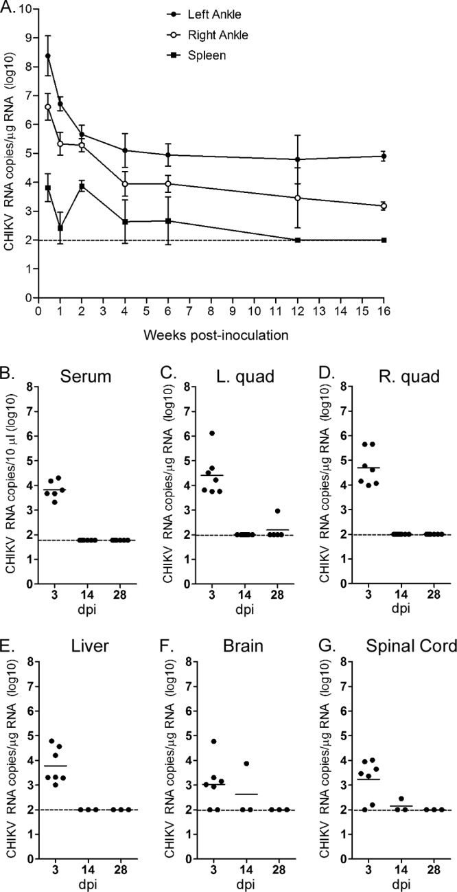
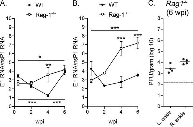
Chikungunya virus (CHIKV) is a reemerging mosquito-borne pathogen that causes incapacitating disease in humans characterized by intense joint pain that can persist for weeks, months, or even years. Although there is some evidence of persistent CHIKV infection in humans suffering from chronic rheumatologic disease symptoms, little is known about chronic disease pathogenesis, and no specific therapies exist for acute or chronic CHIKV disease. To investigate mechanisms of chronic CHIKV-induced disease, we utilized a mouse model and defined the duration of CHIKV infection in tissues and the associated histopathological changes. Although CHIKV RNA was readily detectable in a variety of tissues very early after infection, CHIKV RNA persisted specifically in joint-associated tissues for at least 16 weeks. Inoculation of Rag1(-/-) mice, which lack T and B cells, resulted in higher viral levels in a variety of tissues, suggesting that adaptive immunity controls the tissue specificity and persistence of CHIKV infection. The presence of CHIKV RNA in tissues of wild-type and Rag1(-/-) mice was associated with histopathological evidence of synovitis, arthritis, and tendonitis; thus, CHIKV-induced persistent arthritis is not mediated primarily by adaptive immune responses. Finally, we show that prophylactic administration of CHIKV-specific monoclonal antibodies prevented the establishment of CHIKV persistence, whereas therapeutic administration had tissue-specific efficacy. These findings suggest that chronic musculoskeletal tissue pathology is caused by persistent CHIKV infection and controlled by adaptive immune responses. Our results have significant implications for the development of strategies to mitigate the disease burden associated with CHIKV infection in humans.


Figures

Fig 1
Fig 1
CHIKV RNA persists in joint-associated tissue and spleen of WT mice. Three-week-old WT C57BL/6 mice were mock inoculated (data not shown) or inoculated with 103 PFU of CHIKV by injection in the left rear footpad. Mice were sacrificed and perfused by intracardiac injection with PBS, and total RNA was isolated from the indicated tissues. (A) At 3 dpi (n = 7 mice) and 1 (n = 11), 2 (n = 8), 4 (n = 11), 6 (n = 7), 12 (n = 8), and 16 (n = 3) wpi, CHIKV RNA in the ankles and spleen was quantified by RT-qPCR. Each data point represents the arithmetic mean ± standard errors of the means (SEM), and the dashed line indicates the limit of detection. (B to G) At 3 (n = 7), 14 (n = 3 to 7), and 28 dpi (n = 3 to 5), CHIKV RNA in the serum (B), left quad (C), right quad (D), liver (E), brain (F), and spinal cord (G) was quantified by RT-qPCR. Horizontal lines indicate the means, and dashed lines indicate the limits of detection. Data shown are derived from 2 to 3 independent experiments, except data for 16 wpi, which were derived from a single experiment.
Fig 2
Fig 2
Persistence of CHIKV RNA in joint-associated tissue is virus genotype independent. Three-week-old WT C57BL/6 mice were inoculated with 103 PFU of CHIKV strain 37997 or strain PO731460 by injection in the left rear footpad (n = 6 or 7/group). At 28 dpi, mice were sacrificed and perfused by intracardiac injection with PBS, and total RNA was isolated from the indicated tissues. CHIKV RNA in the left ankle (A) and right ankle (B) was quantified by RT-qPCR. Levels of CHIKV RNA from mice inoculated with 103 PFU of CHIKV strain SL15649 were quantified by the same assay for comparison. Horizontal lines indicate the means, and dashed lines indicate the limits of detection. *, P < 0.05; **, P < 0.01, as determined by one-way ANOVA followed by Tukey's multiple comparison test. Data shown are derived from two independent experiments.
Fig 3
Fig 3
CHIKV persistence in tissues of Rag1−/− mice. Three-week-old Rag1−/− congenic C57BL/6 mice were mock inoculated (data not shown) or inoculated with 103 PFU of CHIKV by injection in the left rear footpad. At 3 dpi (n = 6) and 1 (n = 6), 2 (n = 9), 4 (n = 6), 6 (n = 9), and 12 wpi (n = 5), mice were sacrificed and perfused by intracardial injection with 1× PBS, and total RNA was isolated from the left ankle (A), right ankle (B), left quadriceps muscle (C), right quadriceps muscle (D), and spleen (E). Levels of CHIKV RNA were quantified by RT-qPCR. For comparison, values were plotted against values in WT mice. (F) Serum was analyzed for infectious virus via direct plaque assays. Dashed lines indicate the limit of detection. *, P < 0.05; **, P < 0.01; ***, P < 0.001, as determined by two-way ANOVA followed by Bonferroni posttest analysis. Data shown are derived from at least two independent experiments.
Fig 4
Fig 4
Increased subgenomic mRNA in tissues of Rag1−/− mice. CHIKV RNA in the left ankle (A) and right ankle (B) of WT and Rag1−/− C57BL/6 mice at the indicated time points was quantified by RT-qPCR assays specific for nucleotide sequences in the nsP1 and the E1 genes in parallel as described in Materials and Methods. The E1/nsP1 ratio was plotted as an indirect measurement of subgenomic mRNA production. *, P < 0.05; **, P < 0.01; ***, P < 0.001, as determined by one-way ANOVA followed by Tukey's multiple comparison test. (C) Infectious virus in the left and right ankles of Rag1−/− C57BL/6 mice at 42 dpi was quantified by direct plaque assay. Dashed line indicates the limit of detection. Data shown are derived from at least two independent experiments.
Fig 5
Fig 5
Chronic synovitis in WT and Rag1−/− mice. Three week-old WT and Rag1−/− C57BL/6 mice were mock inoculated or inoculated with 103 PFU of CHIKV by injection in the left rear footpad. (A) At 7 and 42 dpi, 5-μm paraffin-embedded sections were generated from the hind limbs and stained with hematoxylin and eosin. Arrows indicate areas of synovitis, as identified by an anatomic pathologist. Scale bar, 200 μM. Images are representative of three mice per group. (B to F) At 1, 4, 6, and 12 wpi, 5-μm paraffin-embedded sections were generated from the hind limbs, stained with hematoxylin and eosin, and scored in a blinded manner by two anatomic pathologists for the degree of synovitis (B), arthritis (C), metatarsal muscle inflammation (D), metatarsal muscle necrosis (E), and tendonitis (F) based on the following scale for percentage of tissue affected: 0, absent (0%); 1, minimal (<10%); 2, mild (11 to 25%); 3, moderate (26 to 40%); 4, marked (41 to 60%); and 5, severe (>60%).
Fig 6
Fig 6
Efficacy of MAb prophylaxis and therapy against persistent CHIKV infection in Rag1−/− mice. (A to C) Three-week-old Rag1−/− C57BL/6 mice were injected i.p. with 400 μg of WNV E16 MAb or 200 μg CHK-152 and 200 μg CHK-166 MAbs on days −1 and +3. On day 0, mice were inoculated with 103 PFU of CHIKV in the left rear footpad. At 28 dpi, mice were sacrificed and perfused with PBS via intracardiac injection, and infectious virus in the serum (A) was quantified by plaque assay. Tissues (left ankle/foot [B] and right ankle/foot [C]) were homogenized in TRIzol, and CHIKV RNA was quantified by RT-qPCR. Horizontal lines indicate the means, and dashed lines indicate the limits of detection. P values were determined by Mann-Whitney tests. Data are from two independent experiments. (D to G) Three-week-old Rag1−/− C57BL/6 mice were inoculated with 103 PFU of CHIKV in the left rear footpad. On days 21 and 25, mice were injected i.p. with 400 μg of WNV E16 MAb or 200 μg CHK-152 and 200 μg CHK-166 MAbs. At 28 dpi, mice were sacrificed and perfused with PBS via intracardiac injection. (D) Infectious virus in the serum was quantified by plaque assay. The left ankle (E) and right ankle (F) were homogenized in TRIzol, and CHIKV RNA was quantified by RT-qPCR. (G) CHIKV RNA in the right ankle was quantified by RT-qPCR assays specific for nucleotide sequences in the nsP1 and the E1 genes in parallel as described in Materials and Methods. The E1/nsP1 ratio was plotted as an indirect measurement of subgenomic mRNA production. Horizontal bars indicate the means, and dashed lines indicate the limits of detection. P values were determined by the Mann-Whitney test (D) or two-tailed unpaired t tests (F and G). Data are from two independent experiments.

References

    1. Suhrbier A, Jaffar-Bandjee MC, Gasque P. 2012. Arthritogenic alphaviruses—an overview. Nat. Rev. Rheumatol. 8:420–429 - PubMed
    1. Burt FJ, Rolph MS, Rulli NE, Mahalingam S, Heise MT. 2012. Chikungunya: a re-emerging virus. Lancet 379:662–671 - PubMed
    1. Dupuis-Maguiraga L, Noret M, Brun S, Le Grand R, Gras G, Roques P. 2012. Chikungunya disease: infection-associated markers from the acute to the chronic phase of arbovirus-induced arthralgia. PLoS Negl. Trop. Dis. 6:e1446.10.1371/journal.pntd.0001446 - DOI - PMC - PubMed
    1. Ross RW. 1956. The Newala epidemic. III. The virus: isolation, pathogenic properties and relationship to the epidemic. J. Hyg. 54:177–191 - PMC - PubMed
    1. Powers AM, Logue CH. 2007. Changing patterns of Chikungunya virus: re-emergence of a zoonotic arbovirus. J. Gen. Virol. 88:2363–2377 - PubMed

Publication types

MeSH terms

Substances