. 2014 Jan;88(1):110-9.
doi: 10.1128/JVI.02555-13. Epub 2013 Oct 16.

Elucidation of the block to herpes simplex virus egress in the absence of tegument protein UL16 reveals a novel interaction with VP22

Affiliations

Elucidation of the block to herpes simplex virus egress in the absence of tegument protein UL16 reveals a novel interaction with VP22

Jason L Starkey et al. J Virol. 2014 Jan.

Abstract

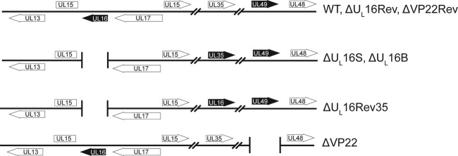
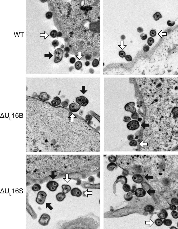
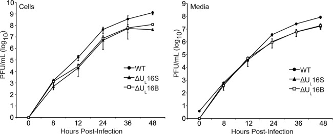
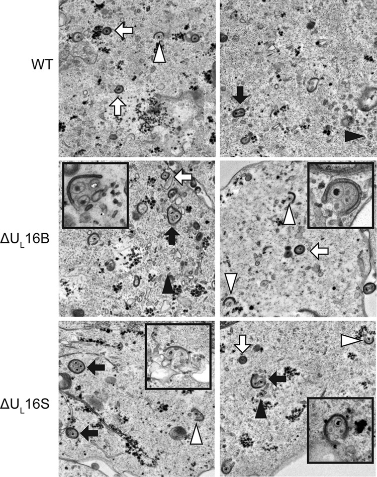
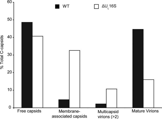
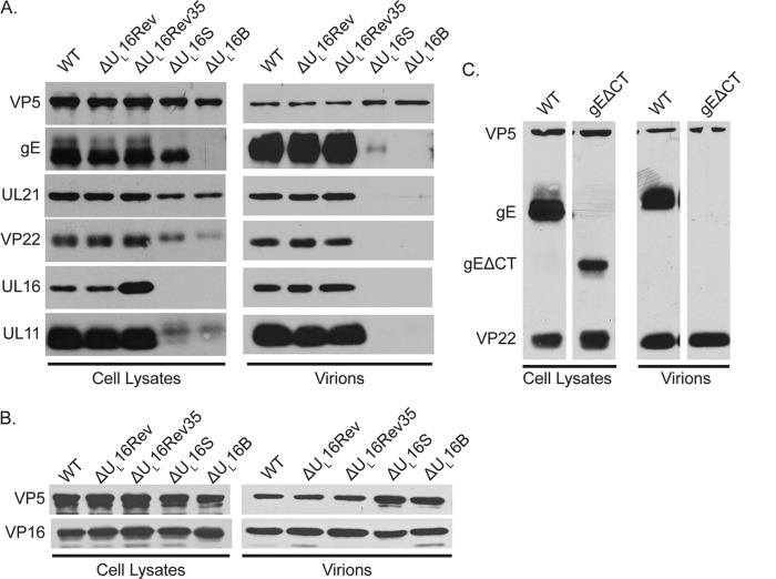
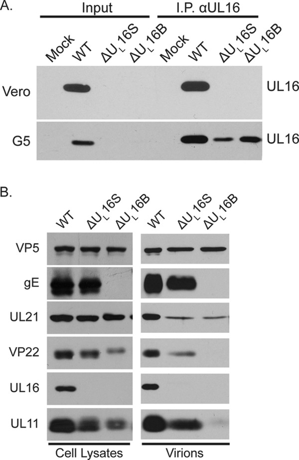
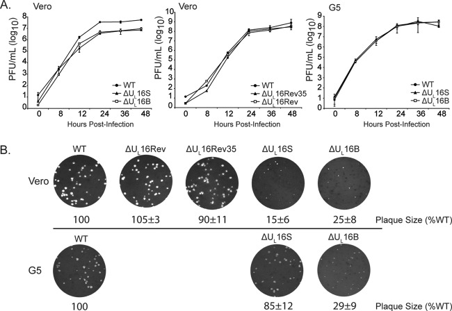
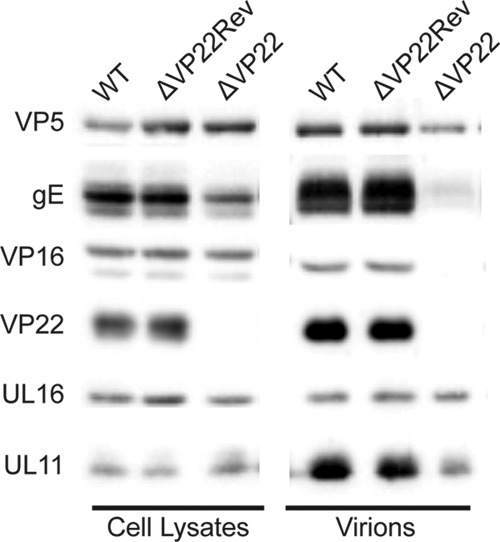
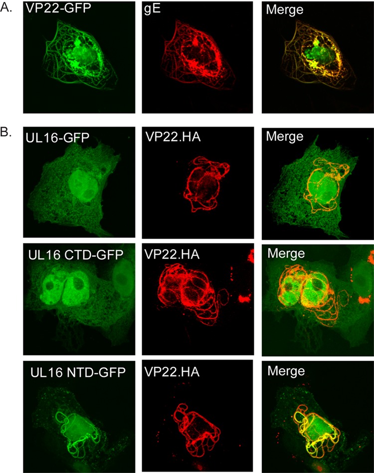
UL16 is a tegument protein of herpes simplex virus (HSV) that is conserved among all members of the Herpesviridae, but its function is poorly understood. Previous studies revealed that UL16 is associated with capsids in the cytoplasm and interacts with the membrane protein UL11, which suggested a "bridging" function during cytoplasmic envelopment, but this conjecture has not been tested. To gain further insight, cells infected with UL16-null mutants were examined by electron microscopy. No defects in the transport of capsids to cytoplasmic membranes were observed, but the wrapping of capsids with membranes was delayed. Moreover, clusters of cytoplasmic capsids were often observed, but only near membranes, where they were wrapped to produce multiple capsids within a single envelope. Normal virion production was restored when UL16 was expressed either by complementing cells or from a novel position in the HSV genome. When the composition of the UL16-null viruses was analyzed, a reduction in the packaging of glycoprotein E (gE) was observed, which was not surprising, since it has been reported that UL16 interacts with this glycoprotein. However, levels of the tegument protein VP22 were also dramatically reduced in virions, even though this gE-binding protein has been shown not to depend on its membrane partner for packaging. Cotransfection experiments revealed that UL16 and VP22 can interact in the absence of other viral proteins. These results extend the UL16 interaction network beyond its previously identified binding partners to include VP22 and provide evidence that UL16 plays an important function at the membrane during virion production.


Figures

FIG 1
FIG 1
Virus mutants. The relevant regions of the HSV-1 genome are shown. Black arrows represent altered genes. The ΔUL16S and ΔVP22 null mutants were generated by removal of their coding sequences (UL16 and UL49, respectively) from the wild-type BAC.KOS plasmid. The UL16 and VP22 coding sequences were restored to generate ΔUL16Rev and ΔVP22Rev, respectively. For the ΔUL16Rev35 strain, the UL35 coding sequence was replaced with that for UL16 in order to rule out context-specific defects associated with the deletion.
FIG 2
FIG 2
Ultrastructural properties of extracellular ΔUL16 virions. Vero cells were infected with the indicated viruses (MOI, 1) 24 h prior to fixing and processing for thin-section electron microscopy. Examples of multicapsid virions (black arrows) and single-capsid virions (white arrows) are indicated.
FIG 3
FIG 3
Growth kinetics of ΔUL16 viruses. Intracellular (Cells) and extracellular (Media) viruses were harvested at the indicated times after infection of Vero cells (MOI, 1), and titers were determined by plaque assays on Vero cells. Each measurement was made in triplicate, and the error bars represent the standard errors of the means.
FIG 4
FIG 4
Multiple capsids are wrapped at once. Representative thin-section electron micrographs of WT- and ΔUL16 mutant-infected (MOI, 1) Vero cells are shown at 24 h postinfection. Simultaneous envelopment of several capsids at a time was detected in ΔUL16 mutant-infected Vero cells (insets). Examples of fully wrapped multicapsid virions (black arrows), single-capsid virions (white arrows), partially wrapped capsids (white arrowheads), and free capsids (black arrowheads) are indicated.
FIG 5
FIG 5
Quantitation of the various species of intracellular capsids. Electron micrographs of Vero cells infected for 24 h with WT or ΔUL16S virus (MOI, 1) were obtained, and the DNA-filled capsids were counted and classified as either free capsids (not near membranes), membrane-associated capsids, multicapsid virions (2 or more capsids fully wrapped with a single envelope), or mature virions (completely wrapped with a single capsid). Micrographs from 3 independent experiments were used, yielding a total of 1,008 WT capsids and 607 ΔUL16S capsids.
FIG 6
FIG 6
Cellular expression and packaging of viral proteins. Vero cells were infected with the indicated viruses at an MOI of 5, and the cultures were harvested 18 to 24 h postinfection. Infected cells were directly dissolved in sample buffer (left side of each panel), while extracellular virions were first concentrated by pelleting through a 30% sucrose cushion and then dissolved in sample buffer (right side of each panel). The samples were analyzed by Western blotting with antibodies against the indicated viral proteins, and the amount of each sample loaded was normalized based on the amount of the major capsid protein, VP5. Blots from one of three independent experiments are shown. (A and B) Results for the ΔUL16 mutants and revertant viruses. (C) Results for the mutant lacking the cytoplasmic tail of gE (gEΔCT).
FIG 7
FIG 7
Expression and packaging of viral proteins in complementing G5 cells. (A) Vero and G5 cells were infected with the indicated viruses, and cytoplasmic lysates were prepared 18 h postinfection. (Input lanes) A fraction of the total lysates was loaded as a control for protein expression. (I.P. lanes) Antibodies were used to immunoprecipitate UL16 and to subsequently detect UL16 expression by Western blot analysis. (B) Viral protein expression and packaging by UL16-deficient viruses in G5 cells.
FIG 8
FIG 8
Growth properties of complemented ΔUL16 viruses. (A) Cultures of Vero or G5 cells were infected with the indicated viruses at an MOI of 1. At various times after infection, the total amount of virus present in the cells and medium (combined) was measured by plaque assays on Vero cells. Measurements from three independent experiments were made, and the error bars represent standard errors of the means. (B) Vero or G5 cells were infected with dilutions of the indicated viruses and were overlaid with methylcellulose. Four days postinfection, the cells were fixed and stained, and plaque sizes relative to those of the wild-type virus were measured. Representative plates from three independent experiments are shown.
FIG 9
FIG 9
Expression and packaging of viral proteins by the VP22-null virus. Vero cells were infected with the indicated viruses (MOI, 5). The cultures were harvested 18 to 24 h postinfection, and the indicated viral proteins present in total-cell lysates (left) and virions (right) were detected by Western blotting.
FIG 10
FIG 10
Colocalization analysis of UL16 and VP22. (A) Vero cells were cotransfected with plasmids expressing VP22-GFP and its binding partner gE as a positive control. (B) Vero cells were cotransfected with HA-tagged, full-length VP22 and the indicated GFP-tagged UL16 constructs. All samples were viewed and imaged by confocal microscopy.

Similar articles

Cited by

References

    1. Johnson DC, Baines JD. 2011. Herpesviruses remodel host membranes for virus egress. Nat. Rev. Microbiol. 9:382–394. 10.1038/nrmicro2559 - DOI - PubMed
    1. Kelly BJ, Fraefel C, Cunningham AL, Diefenbach RJ. 2009. Functional roles of the tegument proteins of herpes simplex virus type 1. Virus Res. 145:173–186. 10.1016/j.virusres.2009.07.007 - DOI - PubMed
    1. Mettenleiter TC, Klupp BG, Granzow H. 2009. Herpesvirus assembly: an update. Virus Res. 143:222–234. 10.1016/j.virusres.2009.03.018 - DOI - PubMed
    1. Han J, Chadha P, Starkey JL, Wills JW. 2012. Function of glycoprotein E of herpes simplex virus requires coordinated assembly of three tegument proteins on its cytoplasmic tail. Proc. Natl. Acad. Sci. U. S. A. 109:19798–19803. 10.1073/pnas.1212900109 - DOI - PMC - PubMed
    1. Harper AL, Meckes DG, Jr, Marsh JA, Ward MD, Yeh PC, Baird NL, Wilson CB, Semmes OJ, Wills JW. 2010. Interaction domains of the UL16 and UL21 tegument proteins of herpes simplex virus. J. Virol. 84:2963–2971. 10.1128/JVI.02015-09 - DOI - PMC - PubMed

Publication types

Substances

LinkOut - more resources