. 2014 Jan;88(1):21-40.
doi: 10.1128/JVI.01558-13. Epub 2013 Oct 16.

The selection of low envelope glycoprotein reactivity to soluble CD4 and cold during simian-human immunodeficiency virus infection of rhesus macaques

Affiliations

The selection of low envelope glycoprotein reactivity to soluble CD4 and cold during simian-human immunodeficiency virus infection of rhesus macaques

Kathleen McGee et al. J Virol. 2014 Jan.

Abstract

Envelope glycoprotein (Env) reactivity (ER) describes the propensity of human immunodeficiency virus type 1 (HIV-1) Env to change conformation from the metastable unliganded state in response to the binding of ligands (antibodies and soluble CD4 [sCD4]) or incubation in the cold. To investigate Env properties that favor in vivo persistence, we inoculated rhesus macaques with three closely related CCR5-tropic simian-human immunodeficiency viruses (SHIVs) that differ in ER to cold (ERcold) and ER to sCD4 (ERsCD4); these SHIVs were neutralized by antibodies equivalently and thus were similar in ERantibody. All three SHIVs achieved high levels of acute viremia in the monkeys without alteration of their Env sequences, indicating that neither ERcold nor ERsCD4 significantly influences the establishment of infection. Between 14 and 100 days following infection, viruses with high ERcold and ERsCD4 were counterselected. Remarkably, the virus variant with low ERcold and low ERsCD4 did not elicit a neutralizing antibody response against the infecting virus, despite the generation of high levels of anti-Env antibodies in the infected monkeys. All viruses that achieved persistent viremia escaped from any autologous neutralizing antibodies and exhibited low ERcold and low ERsCD4. One set of gp120 changes determined the decrease in ERcold and ERsCD4, and a different set of gp120 changes determined resistance to autologous neutralizing antibodies. Each set of changes contributed to a reduction in Env-mediated entry. During infection of monkeys, any Env replication fitness costs associated with decreases in ERcold and ERsCD4 may be offset by minimizing the elicitation of autologous neutralizing antibodies.


Figures

FIG 1
FIG 1
Infectivity and ER-related properties of SHIV Envs. All virus preparations were normalized by reverse transcriptase (RT) activity and used to test infectivity and sensitivity to inhibition. (A) Infectivity of KY, KA, and KY62 Env-pseudotyped reporter viruses was tested in CD4+ CCR5+ Cf2Th cells. The Cf2Th CD4+ CCR5+ cells were lysed 48 h after infection and assayed for luciferase activity. All values are expressed as percent infection at a given virus level compared to the value observed for the maximum input of the KY virus. (B) The infectivity of full-length SHIVs was tested in PBMC pooled from three rhesus macaques. Cell supernatants were collected at the times indicated and assayed for RT activity. (C to F) Cold inactivation (C and E), sCD4 inhibition (D and E), and neutralization sensitivity (F) experiments were performed by using reporter viruses pseudotyped with the KB9, KY, KA, or KY62 Envs. (C) Single-round reporter viruses were incubated on ice for 0 to 24 h before infection. (D and F) Viruses were preincubated at 37°C with 0 to 20 μg/ml of sCD4 (D) or with 10 μg/ml of 2G12, b12, 39F, 17b, 4E10, or VRC01 or 5 μg/ml of PGT128 (F) 1 h before infection. All infections were performed in triplicate with Cf2Th CD4+ CCR5+ target cells, using RT-normalized levels of input virus. Cells were lysed after 48 h and evaluated for luciferase activity. Error bars represent standard errors of the means from two separate experiments. (E) sCD4 reactivity and cold sensitivity of the indicated SHIV Envs are shown. sCD4 reactivity was calculated as described in Materials and Methods and normalized to the ERsCD4 of viruses pseudotyped with KY Env, which was set at a value of 1. Cold sensitivity was calculated as the reciprocal of the time (IT50) on ice required to inactivate 50% of the viral infectivity.
FIG 2
FIG 2
Env determinants of differences in cold sensitivity between SHIV-89.6 and SHIV-KB9. (A and B) Residues in 89.6 Env that differ from those in KB9 Env were altered, alone or in combination, to those found in KB9 Env and vice versa. Recombinant luciferase-expressing HIV-1 pseudotyped with the designated Envs was incubated on ice for the indicated times, and infectivity was determined following incubation with Cf2Th CD4+ CCR5+ cells. Experiments were performed as indicated in the Fig. 1 legend. (C) The approximate ERcold and ERsCD4 values of the SHIV Envs used in this study are plotted. The red arrows indicate phenotypic changes that resulted from modifications of Env introduced in the laboratory. The green arrows indicate phenotypic changes that arose during passage of the virus in monkeys. Note that the ERantibody of all the studied Envs is low.
FIG 3
FIG 3
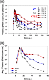
Viremia in SHIV-infected monkeys. (A) Plasma virus levels in rhesus macaques intravenously inoculated with SHIV-KA, SHIV-KY, and SHIV-KY62. (B) Average viremia levels in SHIV-infected monkeys.
FIG 4
FIG 4
Generation of anti-Env antibodies in SHIV-infected monkeys. (A) Levels of antibodies in the plasma of SHIV-infected monkeys that recognize Env of the inoculated SHIV. (B to F) Reporter viruses pseudotyped with the indicated Env were preincubated for 1 h at 37°C with a single dilution of autologous or heterologous plasma (1:50) from the indicated times postinfection. After 1 h at 37°C, the virus-plasma mixtures were incubated with CD4+ CCR5+ Cf2Th cells. Luciferase activity was measured in the target cells. Residual infection is shown as the percentage of reporter activity observed with day 0 (uninfected) plasma. Error bars represent the standard errors of the means.
FIG 5
FIG 5
ERcold, ERsCD4, infectivity, and autologous neutralization of envelope glycoprotein isolates. Env variants derived from SHIV-KY- or -KY62-infected monkeys at the indicated times after inoculation were tested for infectivity and sensitivity to inhibition by incubation of pseudotyped reporter viruses on ice or with inhibitors (sCD4 and contemporaneous plasma). The viruses were then added to Cf2Th CD4+ CCR5+ cells, and luciferase activity was measured 48 h later. (A) Cold sensitivity is shown as the reciprocal of the average time on ice required for a 50% reduction in virus infectivity (IT50). (B) Average soluble CD4 reactivity was calculated as described in Materials and Methods. (C) Average infectivity of variants was calculated from infectivity of maximum virus input levels relative to KY virus infectivity observed with the same RT-normalized virus input. Each experiment was performed with four virus dilutions to ensure that subsaturating infection concentrations were compared. (D) Average autologous neutralization of variants was performed by measuring inhibition of infectivity in the presence of 2% contemporaneous plasma compared to preinfection plasma from the same monkey. All experiments were performed as described in the Fig. 1 legend.
FIG 6
FIG 6
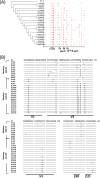
Phylogeny and alignment of SHIV-KY and -KY62 Env sequences. (A) Cladogram (left) and Highlighter analysis (right) of env sequences from animal 354-05 showing synonymous (green) and nonsynonymous (red) mutations. Relative positions of gp120 variable regions are shown, and the gp120/gp41 cleavage point is delineated. (B and C) Alignment of Env sequences spanning the V1/V2, V3, and C4 gp120 regions from SHIV-KY-infected animals 228-91 (days 70 and 98) and 354-05 (days 70 to 434) (B) or SHIV-KY62-infected animals 331-08 and 401-08 (days 70 to 322) (C). Phylogenic tree construction was calculated with PhyML 3.0 (ATCG) (102). Alignments were generated by using Highlighter and SeqPublish (Los Alamos National Laboratory). All amino acid positions are numbered according to the HXB2 HIV-1 prototype.
FIG 6
FIG 6
Phylogeny and alignment of SHIV-KY and -KY62 Env sequences. (A) Cladogram (left) and Highlighter analysis (right) of env sequences from animal 354-05 showing synonymous (green) and nonsynonymous (red) mutations. Relative positions of gp120 variable regions are shown, and the gp120/gp41 cleavage point is delineated. (B and C) Alignment of Env sequences spanning the V1/V2, V3, and C4 gp120 regions from SHIV-KY-infected animals 228-91 (days 70 and 98) and 354-05 (days 70 to 434) (B) or SHIV-KY62-infected animals 331-08 and 401-08 (days 70 to 322) (C). Phylogenic tree construction was calculated with PhyML 3.0 (ATCG) (102). Alignments were generated by using Highlighter and SeqPublish (Los Alamos National Laboratory). All amino acid positions are numbered according to the HXB2 HIV-1 prototype.
FIG 7
FIG 7
Env mutant infectivity, autologous neutralization, ERcold, and ERsCD4. Viruses pseudotyped with KY (left) or d70C (right) Env recombinants were evaluated for infectivity and inhibition by plasma antibodies, cold, and sCD4 relative to the values observed for viruses pseudotyped with wild-type KY or d70C Envs, respectively. (A) Relative infectivity of the viruses pseudotyped with the indicated Env variants. (B) Neutralization of the recombinant viruses by monkey 354-05 plasma from days 0 to 70 p.i. (C and D) Cold sensitivity (C) and sCD4 reactivity (D) of the recombinants are shown. A summary of the KY and d70C Env phenotypes can be found in Tables 2 and 3, respectively. All experiments were performed as described in the Fig. 1 legend. The error bars indicate the standard errors of the means.
FIG 8
FIG 8
Early mutant infectivity, autologous neutralization, ERcold, and ERsCD4. (A and C) Viruses pseudotyped with d70A or d70B Env mutants were evaluated for infectivity, relative to the values observed for wild-type d70A or d70B Envs, respectively. (B and D) Sensitivity of the pseudotyped viruses to neutralization by autologous plasma from days 0 to 70 is shown. (E and F) d70B mutants were also tested for cold sensitivity (E) and sCD4 reactivity (F). All experiments were performed as described in the Fig. 1 legend. Standard errors of the means are indicated by error bars.
FIG 9
FIG 9
Changes in ERcold and ERsCD4 in late virus variants with L317W and/or T440A Env changes. The W317 and/or A440 residue was introduced into d266 Env or was changed to L317 and/or T440 in d434 Env from monkey 354-05. Cold inactivation and sCD4 reactivity of the pseudotyped viruses were measured as described in the Fig. 1 legend. A summary of the phenotypes, including relative infectivity, cold sensitivity, and sCD4 reactivity, for each late variant is described in Table 4.
FIG 10
FIG 10
Energy landscape of HIV-1 Env variants. The metastable, unliganded state of the HIV-1 Env glycoproteins exists in a local energy well. The heights of the activation barriers surrounding this local energy well are inversely related to the ER, ERantibody, ERsCD4, and ERcold (64). Changes in the energy of the metastable unliganded state (asterisk) result in global changes in ER, which in turn result in changes in ERantibody, ERsCD4, and ERcold. Note that “notches” in the hills surrounding the energy well allow ERantibody, ERsCD4, and ERcold to be independently regulated from each other and from ER. ERcold and ERsCD4 often correlate in HIV-1 Env variants (64). The activation energy associated with the binding of target cell CD4 can influence the level of CD4 required on the cell for HIV-1 entry. At the time of seroconversion in SHIV-infected monkeys, HIV-1 Envs with more shallow notches are apparently selected, lowering ERcold, ERsCD4, and entry efficiency. This is predicted to result in an Env that remains longer in the metastable, unliganded state.

Similar articles

Cited by

References

    1. Barre-Sinoussi F, Chermann JC, Rey F, Nugeyre MT, Chamaret S, Gruest J, Dauguet C, Axler-Blin C, Vezinet-Brun F, Rouzioux C, Rozenbaum W, Montagnier L. 1983. Isolation of a T-lymphotropic retrovirus from a patient at risk for acquired immune deficiency syndrome (AIDS). Science 220:868–871. 10.1126/science.6189183 - DOI - PubMed
    1. Gao F, Morrison SG, Robertson DL, Thornton CL, Craig S, Karlsson G, Sodroski J, Morgado M, Galvao-Castro B, von Briesen H, Beddows S, Weber J, Sharp PM, Shaw GM, Hahn BH. 1996. Molecular cloning and analysis of functional envelope genes from human immunodeficiency virus type 1 sequence subtypes A through G. The WHO and NIAID networks for HIV isolation and characterization. J. Virol. 70:1651–1667 - PMC - PubMed
    1. Robey WG, Safai B, Oroszlan S, Arthur LO, Gonda MA, Gallo RC, Fischinger PJ. 1985. Characterization of envelope and core structural gene products of HTLV-III with sera from AIDS patients. Science 228:593–595. 10.1126/science.2984774 - DOI - PubMed
    1. Veronese FD, DeVico AL, Copeland TD, Oroszlan S, Gallo RC, Sarngadharan MG. 1985. Characterization of gp41 as the transmembrane protein coded by the HTLV-III/LAV envelope gene. Science 229:1402–1405. 10.1126/science.2994223 - DOI - PubMed
    1. Wyatt R, Sodroski J. 1998. The HIV-1 envelope glycoproteins: fusogens, antigens, and immunogens. Science 280:1884–1888. 10.1126/science.280.5371.1884 - DOI - PubMed

Publication types

LinkOut - more resources