

Secreted proteins from carotid endarterectomy: an untargeted approach to disclose molecular clues of plaque progression
- PMID: 24131807
- PMCID: PMC3853772
- DOI: 10.1186/1479-5876-11-260
Secreted proteins from carotid endarterectomy: an untargeted approach to disclose molecular clues of plaque progression
Abstract
Background: Atherosclerosis is the main cause of morbidity and mortality in Western countries and carotid plaque rupture is associated to acute events and responsible of 15-20% of all ischemic strokes. Several proteomics approaches have been up to now used to elucidate the molecular mechanisms involved in plaque formation as well as to identify markers of pathology severity for early diagnosis or target of therapy. The aim of this study was to characterize the plaque secretome. The advantage of this approach is that secretome mimics the in vivo condition and implies a reduced complexity compared to the whole tissue proteomics allowing the detection of under-represented potential biomarkers.
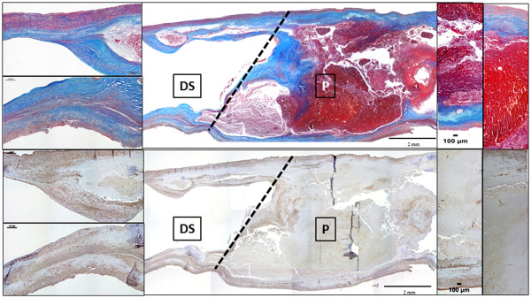
Methods: Secretomes from carotid endarterectomy specimens of 14 patients were analyzed by a liquid chromatography approach coupled with label free mass spectrometry. Differential expression of proteins released from plaques and from their downstream distal side segments were evaluated in each specimen. Results were validated by Western blot analysis and ELISA assays. Histology and immunohistochemistry were performed to characterize plaques and to localise the molecular factors highlighted by proteomics.
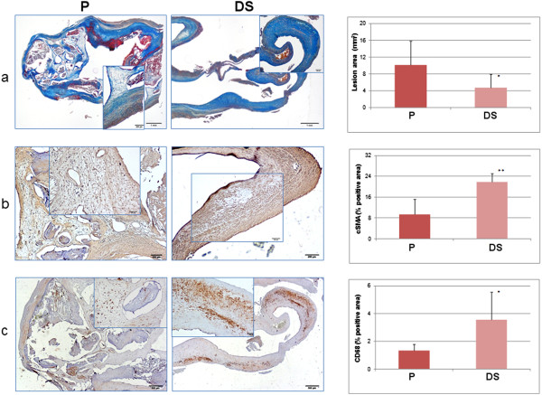
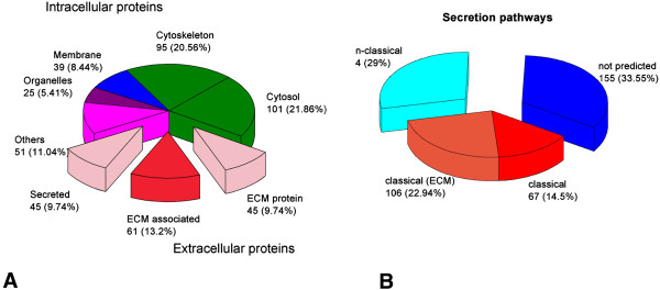
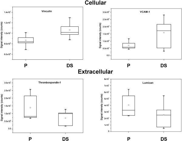
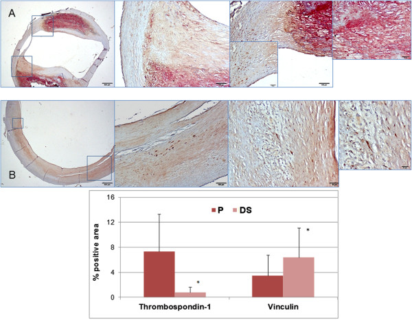
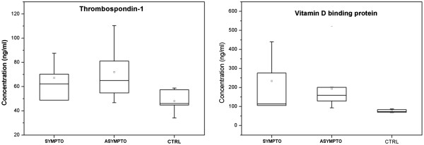
Results: A total of 463 proteins were identified and 31 proteins resulted differentially secreted from plaques and corresponding downstream segments. A clear-cut distinction in the distribution of cellular- and extracellular-derived proteins, evidently related to the higher cellularity of distal side segments, was observed along the longitudinal axis of carotid endarterectomy samples. The expressions of thrombospondin-1, vitamin D binding protein, and vinculin, as examples of extracellular and intracellular proteins, were immunohistologically compared between adjacent segments and validated by antibody assays. ELISA assays of plasma samples from 34 patients and 10 healthy volunteers confirmed a significantly higher concentration of thrombospondin-1 and vitamin D binding protein in atherosclerotic subjects.
Conclusions: Taking advantage of the optimized workflow, a detailed protein profile related to carotid plaque secretome has been produced which may assist and improve biomarker discovery of molecular factors in blood. Distinctive signatures of proteins secreted by adjacent segments of carotid plaques were evidenced and they may help discriminating markers of plaque complication from those of plaque growth.
Figures

Similar articles
-
Extracellular matrix proteomics identifies molecular signature of symptomatic carotid plaques.J Clin Invest. 2017 Apr 3;127(4):1546-1560. doi: 10.1172/JCI86924. Epub 2017 Mar 20. J Clin Invest. 2017. PMID: 28319050 Free PMC article.
-
Extracellular matrix characterization in plaques from carotid endarterectomy by a proteomics approach.Talanta. 2017 Nov 1;174:341-346. doi: 10.1016/j.talanta.2017.06.014. Epub 2017 Jun 10. Talanta. 2017. PMID: 28738590
-
The relationship with the stability between GRP78, CHOP and human carotid atherosclerotic plaque.Clin Neurol Neurosurg. 2022 Jan;212:107067. doi: 10.1016/j.clineuro.2021.107067. Epub 2021 Nov 25. Clin Neurol Neurosurg. 2022. PMID: 34839153
-
A systemic review into carotid plaque features as predictors of restenosis after carotid endarterectomy.J Vasc Surg. 2021 Jun;73(6):2179-2188.e4. doi: 10.1016/j.jvs.2020.10.084. Epub 2020 Nov 27. J Vasc Surg. 2021. PMID: 33253876
-
Immune cells in carotid artery plaques: what can we learn from endarterectomy specimens?Int Angiol. 2020 Feb;39(1):37-49. doi: 10.23736/S0392-9590.19.04250-0. Epub 2019 Nov 25. Int Angiol. 2020. PMID: 31782285 Review.
Cited by
-
Novel Multiomics Profiling of Human Carotid Atherosclerotic Plaques and Plasma Reveals Biliverdin Reductase B as a Marker of Intraplaque Hemorrhage.JACC Basic Transl Sci. 2018 Aug 1;3(4):464-480. doi: 10.1016/j.jacbts.2018.04.001. eCollection 2018 Aug. JACC Basic Transl Sci. 2018. PMID: 30175270 Free PMC article.
-
Proteomic Studies of Blood and Vascular Wall in Atherosclerosis.Int J Mol Sci. 2021 Dec 9;22(24):13267. doi: 10.3390/ijms222413267. Int J Mol Sci. 2021. PMID: 34948066 Free PMC article. Review.
-
Site-Specific Secretome Map Evidences VSMC-Related Markers of Coronary Atherosclerosis Grade and Extent in the Hypercholesterolemic Swine.Dis Markers. 2015;2015:465242. doi: 10.1155/2015/465242. Epub 2015 Aug 25. Dis Markers. 2015. PMID: 26379359 Free PMC article.
-
Search for Reliable Circulating Biomarkers to Predict Carotid Plaque Vulnerability.Int J Mol Sci. 2020 Nov 3;21(21):8236. doi: 10.3390/ijms21218236. Int J Mol Sci. 2020. PMID: 33153204 Free PMC article. Review.
-
Inflammation blood and tissue factors of plaque growth in an experimental model evidenced by a systems approach.Front Genet. 2014 Apr 7;5:70. doi: 10.3389/fgene.2014.00070. eCollection 2014. Front Genet. 2014. PMID: 24778640 Free PMC article.
References
-
- Touzé E, Mas JL, Röther J, Goto S, Hirsch AT, Ikeda Y, Liau C-S, Ohman EM, Richard AJ, Wilson PWF, Steg PG, Bhatt DL. Impact of carotid endarterectomy on medical secondary prevention after a stroke or a transient ischemic attack: results from the Reduction of Atherothrombosis for Continued Health (REACH) registry. Stroke. 2006;37:2880–2885. doi: 10.1161/01.STR.0000249411.44097.5b. - DOI - PubMed
-
- Puig O, Yuan J, Stepaniants S, Zieba R, Zycband E, Morris M, Coulter S, Yu X, Menke J, Woods J, Chen F, Ramey DR, He X, O'Neill EA, Hailman E, Jhons DG, Hubbard BK, Lum PY, Wright SD, DeSouza MM, Plump A, Reiser W. Gene expression signature that classifies human atherosclerotic plaque by relative inflammation status. Circ Cardiovasc Genet. 2011;4:595–604. doi: 10.1161/CIRCGENETICS.111.960773. - DOI - PubMed
Publication types
MeSH terms
Substances
LinkOut - more resources
Full Text Sources
Other Literature Sources
